
Adrenochrome is what adrenaline turns into when it oxidises. The reaction happens in a beaker within minutes. Its red-purple product has a real and unglamorous job in the laboratory, as the readout in the standard superoxide dismutase assay.
Almost everything else attached to the name traces to one hypothesis published in 1952, which failed. A historian of psychology later concluded its central argument was circular [4].
The interesting chemistry sits somewhere else entirely. Adrenochrome belongs to a family of catecholamine oxidation products called aminochromes. Its dopamine-derived cousin has become a serious candidate for the trigger of Parkinson’s disease [7].
Chemical identity
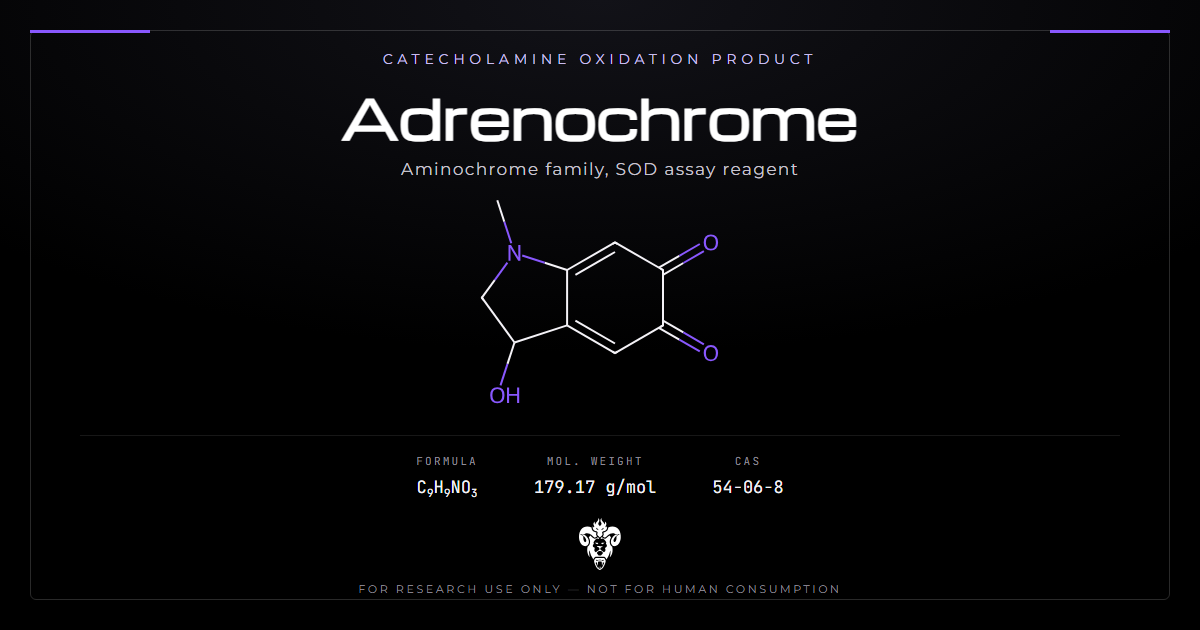
An indoline ring bearing two ketones on the six-membered ring, formed when adrenaline oxidises and cyclises.
| Property | Value |
|---|---|
| Systematic name | 3-hydroxy-1-methyl-2,3-dihydroindole-5,6-dione |
| Common names | Adrenochrome, adraxone |
| Molecular formula | C9H9NO3 |
| Average mass | 179.17 g/mol |
| Monoisotopic mass | 179.0582 Da |
| CAS number | 54-06-8 |
| PubChem CID | 5898 |
| InChIKey | RPHLQSHHTJORHI-UHFFFAOYSA-N |
| Parent compound | Adrenaline (epinephrine) |
| Chemical class | Ortho-quinone, aminochrome |
| Appearance | Red to purple solid or solution |
The ortho-quinone
Two adjacent carbonyls on an aromatic ring make an ortho-quinone, and that arrangement governs everything the molecule does.
Ortho-quinones are electrophiles. They accept electrons readily and form covalent adducts with thiols and amines on proteins. They also redox cycle, generating reactive oxygen species as they shuttle between quinone and semiquinone states.
Nothing about that reactivity is unusual or mysterious. It is the same chemistry that makes cut apples go brown, running on catecholamines instead of plant phenolics.
How the oxidation proceeds
Three steps separate adrenaline from adrenochrome, and all of them happen without enzymes.
Oxidation of the catechol ring gives an ortho-quinone. That quinone carries a secondary amine on its side chain, positioned to attack the ring, so the molecule closes on itself in an intramolecular addition. The resulting indoline oxidises once more to give the coloured product.
Nothing in that sequence requires a catalyst. Dissolved oxygen and a trace of transition metal are enough. That is why adrenaline solutions discolour on standing, and why the reaction serves as a convenient superoxide source.
The same sequence explains the family resemblance across the aminochromes. Every catecholamine with an amine two carbons from the ring can cyclise the same way.
The stereocentre nobody specifies
Carbon 3 carries a hydroxyl and four different substituents, which makes it a stereocentre.
Adrenaline is chiral, and the natural hormone is the L enantiomer. Oxidation preserves that centre, so material derived from L-adrenaline should be a single enantiomer. PubChem’s record for CID 5898 carries no stereodescriptor at all, and the InChIKey ends in UHFFFAOYSA, the hash for an unspecified structure.
That mismatch is worth knowing before ordering. A certificate quoting the flat InChIKey has said nothing about which enantiomer, or whether the material is racemic.
The aminochrome family
One reaction, several catecholamines, several products with the same core.
| Catecholamine | Oxidation product | Downstream polymer |
|---|---|---|
| Adrenaline | Adrenochrome | Melanin-type pigment |
| Noradrenaline | Noradrenochrome | Neuromelanin |
| Dopamine | Aminochrome (dopaminochrome) | Neuromelanin |
Smythies grouped these together (PMID 8637929). He proposed that neuromelanin exists partly to protect catecholamine neurons from the quinones formed along the way [1]. The aminochromes are intermediates on the path from catecholamine to pigment, and the pigment is the safe endpoint.
That ordering explains an otherwise odd fact. Neuromelanin accumulates across a lifetime in exactly the neurons most vulnerable in Parkinson’s disease, yet the pigment itself is not the problem. What matters is the reactive intermediate passing through on the way to it, and how quickly the cell disposes of that.
That framing matters for reading anything about adrenochrome. Findings on one member do not automatically transfer to another. The neurons that make them, the enzymes that detoxify them and the tissue concentrations all differ.
What laboratories actually use it for
The compound’s steady role in current research is as a coloured product you measure.
The superoxide dismutase assay
Adrenaline autoxidises in alkaline buffer. That reaction generates superoxide radicals and yields adrenochrome, which absorbs strongly in the visible range. Superoxide dismutase removes the superoxide and therefore slows the colour forming.
Measuring how much the enzyme suppresses the colour gives its activity. Sirota describes the reaction as modelling the quinoid pathway of adrenaline conversion, and developed a polarographic version tracking oxygen consumption alongside the spectrophotometric one [6].
The assay turns up constantly under the name “epinephrine method”. Bhosle and colleagues measured superoxide dismutase in cervical carcinoma biopsies before and after radiotherapy with it [11]. The same reaction reads out hydroxyl radical scavenging in natural product screening [3].
As a radical detector
Because adrenaline oxidation depends on radical species, the appearance of adrenochrome reports on their presence.
Isoyan and colleagues used exactly that. They quantified a superoxide-producing lipoprotein fraction from Stevia leaves by its ability to oxidise adrenaline, then expressed the result in units of adrenaline oxidation activity [12].
This is the compound’s honest place in the modern literature. It is a chromophore with a known formation pathway, useful because both halves of that sentence are true.
The serious literature is about its dopamine cousin
Aminochrome, the dopamine-derived member of the family, has become one of the more interesting hypotheses in Parkinson’s research.
Why the dopamine version gets the attention
Dopaminergic neurons of the substantia nigra oxidise dopamine to neuromelanin as a normal process, and aminochrome forms inside those cells as an intermediate [8].
The anatomy is what makes the hypothesis attractive. Parkinson’s disease destroys a specific, pigmented population rather than dopaminergic neurons generally, and any credible trigger has to explain that selectivity. A toxin generated only where the pigment is made does explain it.
Segura-Aguilar and colleagues note the paradox that drives the field. A neurotoxic quinone forms routinely inside these neurons, yet healthy people reach old age with the neurons intact [9]. Something must be handling it, and whatever that is may fail in disease.
What it does to a neuron
The proposal is that aminochrome triggers most of the accepted mechanisms of nigral neurodegeneration rather than sitting downstream of them.
Segura-Aguilar lists five of six (PMID 26695514): mitochondrial dysfunction, protein degradation failure, aggregation of alpha-synuclein into neurotoxic oligomers, oxidative stress with neuroinflammation, and endoplasmic reticulum stress [7].
Herrera and colleagues make the same case. They frame the open question as what initially triggers a process everyone otherwise agrees on [8].
The autophagy work is the most concrete. Muñoz and colleagues report two distinct routes [5]. Aminochrome forms alpha-synuclein protofibrils that inactivate chaperone-mediated autophagy. It also forms adducts with alpha- and beta-tubulin, aggregating the microtubules that autophagosomes need in order to fuse with lysosomes.
The single-neuron model
One property of these quinones shapes the whole hypothesis. Aminochrome is either reduced rapidly by flavoenzymes or forms adducts with nearby proteins, so it cannot travel.
Huenchuguala and Segura-Aguilar build a single-neuron degeneration model on that [10]. A toxin that cannot spread to neighbours would produce exactly the extremely slow, cell-by-cell decline seen before motor symptoms appear.
Why most neurons survive it
Two enzymes explain the paradox, and they are the reason the hypothesis is testable rather than merely plausible.
DT-diaphorase and glutathione transferase M2-2
DT-diaphorase reduces the quinone by two electrons, avoiding the one-electron route that would produce a redox-cycling semiquinone. Glutathione transferase M2-2 conjugates it for disposal.
Both prevent aminochrome neurotoxicity, and Segura-Aguilar and colleagues argue their presence is why melanin-containing dopaminergic neurons stay intact in healthy brains [9]. Astrocytes contribute by secreting exosomes carrying the transferase to neurons.
Smythies had already connected the same enzyme family to the adrenaline version [2]. He noted reports of a defective glutathione S-transferase gene in schizophrenia, alongside the enzyme’s role in detoxifying adrenochrome.
Two electrons or one
The distinction between the two reduction routes is the whole reason one enzyme protects and another would not.
A one-electron reduction gives a semiquinone radical, which reacts with oxygen to regenerate the quinone and release superoxide. That cycle consumes reducing equivalents and produces oxidative stress indefinitely from a small amount of quinone.
DT-diaphorase adds two electrons at once, skipping the radical entirely and giving a hydroquinone that the cell can conjugate and excrete [9]. The enzyme is protective because of which route it takes, not because it is faster.
The therapeutic reading
If the enzymes are protective, raising their expression should help.
Huenchuguala and Segura-Aguilar review natural compounds activating the KEAP1/Nrf2 pathway, which increases expression of both DT-diaphorase and glutathione transferase [10]. That is the current translational proposal, and it remains preclinical.
The 1952 hypothesis
Everything else attached to this compound comes from one line of work in Saskatchewan.
Abram Hoffer and Humphry Osmond proposed in 1952 that adrenochrome, formed endogenously from adrenaline, caused schizophrenia (PMID 20533770). They followed it with reports that large doses of niacin reversed the illness, including a double-blind study [4].
The hypothesis was not fringe when it was made. It belonged to the transmethylation theories then dominating biological psychiatry. Mills places it inside the mainstream psychopharmacology of the period rather than outside it [4].
Reports that adrenochrome produced psychotomimetic effects in volunteers date from the same programme. Smythies reviewed those experiments fifty years later while arguing the hypothesis deserved reconsideration [2].
What happened to that hypothesis
It did not survive.
Mills examined the empirical evidence against it and analysed the diagnostic procedures behind it [4]. Hoffer had created and used his own biochemical marker for schizophrenia.
Mills argues that both the conceptualisation of the illness and the treatment approach rested on a tautology. The marker defined the disease that the marker was then said to detect.
The niacin results did not replicate outside that group. Later work on niacin in schizophrenia treats the adrenochrome proposal as one historical mechanism among several rather than an established one.
The original psychotomimetic reports have the same problem as the rest. They come from one research programme, in a small number of volunteers, without independent replication, and modern work has not revisited them.
Smythies argued in 2002 that adrenochrome might still occur as an adrenaline metabolite in the C2 adrenergic neurons of the medulla [2]. That is a proposal about where a molecule might form, not evidence that it causes an illness.
How to read a study of this compound
Four questions separate a study that says something about adrenochrome from one that does not.
Is the compound the subject or the reagent? Most papers indexed under adrenochrome use it to measure superoxide dismutase [6][11]. Those report on the enzyme, not on the quinone.
Which aminochrome? Findings on the dopamine-derived quinone belong to the Parkinson’s literature and do not transfer by name similarity [7][8].
Was it formed in situ or added? A quinone generated inside a cell during pigment synthesis meets the protective enzymes immediately [9]. Adding it from outside skips that step and tests something different.
What concentration? These compounds form transiently at low levels inside particular neurons. Bath application at micromolar concentrations models nothing physiological, and the papers proposing the neurodegeneration hypothesis are explicit that the endogenous route is what interests them [10].
What the record does not establish
No controlled modern trial has tested adrenochrome for anything in humans. The compound has no approved use, and its clinical file consists of a hypothesis from 1952 and the reports generated by its proponents.
Read-across from aminochrome does not work either. That literature concerns a dopamine-derived quinone forming inside specific neurons, at concentrations nobody administers. Adrenochrome differs in its parent catecholamine, its site of formation and its detoxification profile [1][8].
The enzyme work is preclinical throughout. DT-diaphorase and glutathione transferase protection comes from cell and animal models, and the KEAP1/Nrf2 proposal has not reached controlled human testing [9][10].
Nothing in the literature supports the claims that circulate about this compound outside scientific sources. Those trace to the 1950s psychotomimetic reports by way of popular writing, and the underlying hypothesis was found wanting on its own terms [4].
The stereochemistry is unresolved in the reference databases, so even the identity question is looser than it should be for a compound with a defined natural precursor.
Verifying research material
Instability is the defining practical problem. This is an oxidation product that continues to oxidise.
Why it is hard to keep
Every property that makes the compound useful also makes it short-lived.
Stability
Adrenochrome degrades in solution, faster in alkaline conditions, and polymerises toward melanin-type pigments. A solution that darkens from red toward brown or black has moved on.
Prepare fresh, keep cold and dark, and expect a short working window. Any assay using it as a standard should measure absorbance against a freshly prepared curve rather than a stored one.
The compound also forms adducts with thiols, so buffers containing dithiothreitol, mercaptoethanol or free cysteine will consume it. That reactivity is the whole point of the molecule, and it does not switch off because the experiment would prefer it to.
Trace metals matter for the same reason. Iron and copper catalyse the autoxidation that produces the compound, so a chelator such as EDTA stabilises solutions and glassware carrying metal residues destabilises them.
For assay work the practical consequence is that adrenochrome should generally be generated rather than stored. Running the adrenaline autoxidation fresh each session gives a more reproducible baseline than diluting a stock that has been degrading since it was opened.
Identity
Mass is 179.17 average and 179.0582 monoisotopic. The visible absorbance band near 480 nm is the practical identity and purity check, since that is the band the superoxide dismutase assay reads.
Adrenaline itself is the expected residual starting material, and melanin-type polymers the expected degradation products. Neither is invisible: unreacted adrenaline shows in the ultraviolet, and polymer shows as broad absorbance and eventually as insoluble material.
Ask for the enantiomeric form in writing, given that the common database record specifies none.
Kimera publishes third-party certificates of analysis for every lot in its COA database. Laboratories source adrenochrome as an assay reagent and catecholamine oxidation standard. It pairs with glutathione where transferase-mediated conjugation is the subject, or with NAD+ and CoQ10 where a different redox couple is under study. Related work appears in the redox and cofactors category.
Where the name outran the chemistry
One more thing is worth saying plainly, because it explains why this page exists.
Adrenochrome is searched far more often than its research literature would predict. That gap comes from popular writing of the 1950s and 1960s, which picked up the psychotomimetic reports while they still looked promising, and from later repetition that dropped the scientific context entirely.
The compound underneath is ordinary. It is a catecholamine oxidation product that forms whenever adrenaline sits in an open vial, it serves as a convenient chromophore for measuring an antioxidant enzyme, and its most interesting relative is being investigated as a possible trigger for Parkinson’s disease.
None of the claims that circulate outside the scientific literature have support in it. The hypothesis that generated them was examined and found to rest on circular reasoning [4], and no modern controlled work has revisited the human reports.
Common questions about adrenochrome
What is adrenochrome? The oxidation product of adrenaline, an ortho-quinone of the aminochrome family, C9H9NO3 at 179.17 g/mol. It forms spontaneously when adrenaline solutions oxidise.
What do researchers use it for? Mostly as the coloured readout in the superoxide dismutase assay, where adrenaline autoxidation to adrenochrome is suppressed by the enzyme [6][11].
Is it the same as aminochrome? No, though they belong to one family. Aminochrome comes from dopamine, adrenochrome from adrenaline, and the Parkinson’s literature concerns the dopamine version [7][8].
Does it cause schizophrenia? The 1952 hypothesis proposing that failed. A historical analysis found the diagnostic framework circular and the supporting evidence unreplicated [4].
Why is it red? The ortho-quinone chromophore absorbs in the visible range near 480 nm, which is what makes the assay work.
Why does it need protecting from light and air? It is itself an oxidation product and keeps oxidising, polymerising toward melanin-type pigments. Solutions darken as that proceeds.
Is there a stereochemistry question? Yes. Carbon 3 is a stereocentre inherited from L-adrenaline, but the common PubChem record specifies no configuration, so the enantiomeric form should be requested explicitly.
Summary of the evidence
Identity: 3-hydroxy-1-methyl-2,3-dihydroindole-5,6-dione, C9H9NO3, 179.17 g/mol, CAS 54-06-8. An ortho-quinone formed by oxidation of adrenaline.
Family: one of the aminochromes, alongside noradrenochrome and dopamine-derived aminochrome, all intermediates toward melanin-type pigments [1].
Laboratory role: the chromophore in the superoxide dismutase assay and related radical detection methods [6][11][3][12].
The aminochrome literature: the dopamine-derived quinone triggers five of the six accepted mechanisms of nigral neurodegeneration [7][8]. It disrupts chaperone-mediated autophagy and microtubule function [5], and supports a single-neuron degeneration model because it cannot spread [10].
Protection: DT-diaphorase and glutathione transferase M2-2 prevent aminochrome neurotoxicity, which explains why healthy neurons survive routine exposure [9]. Raising their expression through KEAP1/Nrf2 is the current preclinical proposal [10].
The historical hypothesis: proposed in 1952, promoted with niacin treatment claims, and analysed in 2010 as resting on a tautology with unreplicated support [4][2].
Status: supplied for laboratory research use only.
References
- Smythies J. On the function of neuromelanin. Proc Biol Sci. 1996;263(1369):487-489. PMID 8637929. DOI
- Smythies J. The adrenochrome hypothesis of schizophrenia revisited. Neurotox Res. 2002;4(2):147-150. PMID 12829415. DOI
- Fernández F, Torres M. Evaluation of Pluchea carolinensis extracts as antioxidants by the epinephrine oxidation method. Fitoterapia. 2006;77(3):221-226. PMID 16564648. DOI
- Mills JA. Hallucinogens as hard science: the adrenochrome hypothesis for the biogenesis of schizophrenia. Hist Psychol. 2010;13(2):178-195. PMID 20533770. DOI
- Muñoz P, Huenchuguala S, Paris I, Segura-Aguilar J. Dopamine oxidation and autophagy. Parkinsons Dis. 2012;2012:920953. PMID 22966478. DOI
- Sirota TV. A new approach to studying the autoxidation of adrenaline: possibility of the determination of superoxide dismutase activity and the antioxidant properties of various preparations by polarography. Biomed Khim. 2012;58(1):77-87. PMID 22642154. DOI
- Segura-Aguilar J, Muñoz P, Paris I. Aminochrome as new preclinical model to find new pharmacological treatment that stop the development of Parkinson’s disease. Curr Med Chem. 2016;23(4):346-359. PMID 26695514. DOI
- Herrera A, Muñoz P, Steinbusch HWM, Segura-Aguilar J. Are dopamine oxidation metabolites involved in the loss of dopaminergic neurons in the nigrostriatal system in Parkinson’s disease? ACS Chem Neurosci. 2017;8(4):702-711. PMID 28233992. DOI
- Segura-Aguilar J, Muñoz P, Inzunza J, et al. Neuroprotection against aminochrome neurotoxicity: glutathione transferase M2-2 and DT-diaphorase. Antioxidants (Basel). 2022;11(2):296. PMID 35204179. DOI
- Huenchuguala S, Segura-Aguilar J. Natural compounds that activate the KEAP1/Nrf2 signaling pathway as potential new drugs in the treatment of idiopathic Parkinson’s disease. Antioxidants (Basel). 2024;13(9):1125. PMID 39334784. DOI
- Bhosle SM, Pandey BN, Huilgol NG, Mishra KP. Membrane oxidative damage and apoptosis in cervical carcinoma cells of patients after radiation therapy. Methods Cell Sci. 2002;24(1-3):65-68. PMID 12815293.
- Isoyan AS, Simonyan KV, Simonyan RM, et al. Superoxide-producing lipoprotein fraction from Stevia leaves: definition of specific activity. BMC Complement Altern Med. 2019;19(1):88. PMID 31023287. DOI
Adrenochrome is sold for laboratory research use only. Not for human consumption, nor medical, veterinary, or household uses.
Literature retrieved from PubMed.