
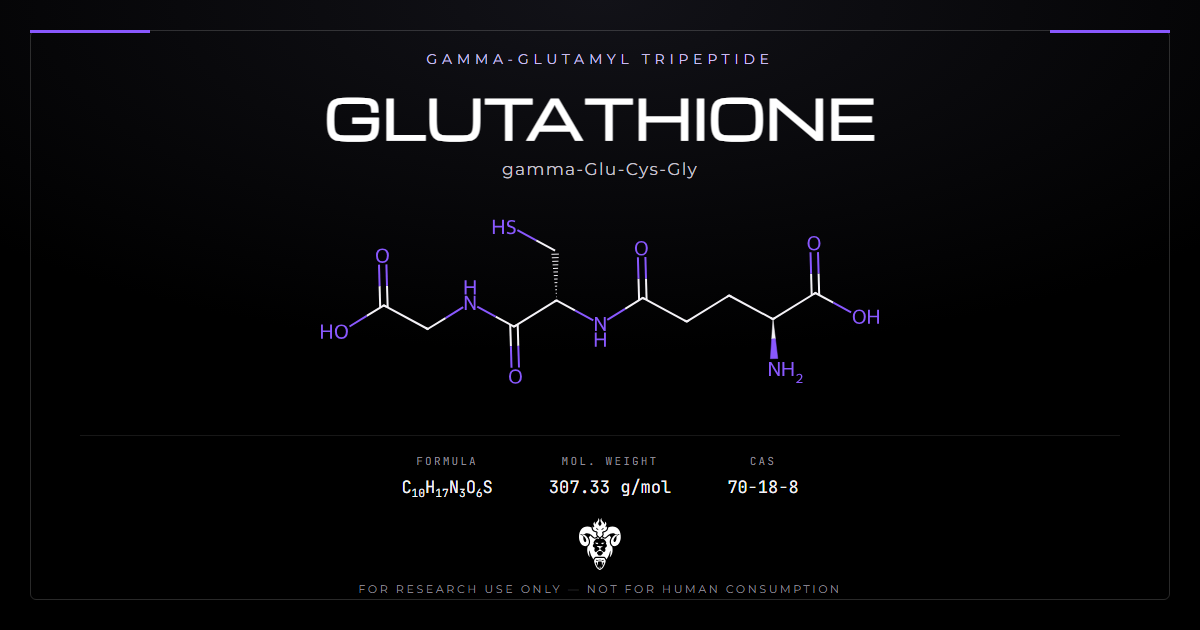
Glutathione is the most abundant thiol inside a cell. The most repeated claim about it is that swallowing it is pointless.
That claim traces to a single study of seven volunteers published in 1992. A six-month randomised controlled trial published in 2014 reached the opposite conclusion. Both are sound, and they answer different questions.
Sorting that out means starting with the chemistry. One unusual bond explains the metabolism, the turnover, the supplementation argument and the analytical difficulties at once.
Chemical identity: a tripeptide with an unusual bond
Glutathione is glutamate, cysteine and glycine. Calling it a tripeptide is accurate and slightly misleading.
| Property | Value |
|---|---|
| Systematic name | (2S)-2-amino-5-[[(2R)-1-(carboxymethylamino)-1-oxo-3-sulfanylpropan-2-yl]amino]-5-oxopentanoic acid |
| Common names | Glutathione, GSH, reduced glutathione |
| Sequence | γ-L-glutamyl-L-cysteinyl-glycine |
| Molecular formula | C10H17N3O6S |
| Molecular weight | 307.33 g/mol |
| CAS number | 70-18-8 |
| PubChem CID | 124886 |
| InChIKey | RWSXRVCMGQZWBV-WDSKDSINSA-N |
| Reactive group | Free thiol on cysteine |
| Oxidised form | Glutathione disulfide (GSSG), 612.6 g/mol |
| Intracellular concentration | Millimolar in most cell types |
The gamma-glutamyl bond
In an ordinary peptide, each residue links through its alpha-carboxyl. In glutathione, glutamate links through its side-chain carboxyl instead. That is the gamma bond, and it is the single most consequential feature of the molecule.
Ordinary peptidases cannot cleave it. One enzyme can. Gamma-glutamyl transpeptidase sits on the outer surface of cell membranes, abundant in intestine, kidney and liver.
Two things follow. The molecule survives inside cells where ordinary tripeptides would be degraded, so concentrations reach millimolar. It is also taken apart at the tissues expressing that one enzyme, which is exactly what the oral dosing argument turns on.
Why the concentration is so high
Millimolar is an unusual concentration for anything that is not a bulk metabolite. Most signalling molecules work at nanomolar, and most peptides never accumulate at all.
Two features make it possible. The gamma linkage blocks the peptidases that would otherwise degrade it, and cells make it continuously in two ATP-dependent steps rather than importing it.
The consequence is that the pool functions as a buffer rather than a signal. A buffer has to be present in large excess over the thing it absorbs, and that is precisely how cells use this one against electrophiles and peroxides.
Reduced and oxidised forms are not interchangeable
The cysteine thiol is the working group. Two glutathione molecules oxidise to one glutathione disulfide, and glutathione reductase reduces it back using NADPH.
Almost the entire intracellular pool sits in the reduced form. The ratio between them reports the redox state of a compartment better than the absolute amount does. Studies use it that way [11][12].
What glutathione does in a cell
Three distinct jobs, often conflated into one word.
The redox buffer
The GSH/GSSG couple is the principal redox buffer of the cytosol. The pool is large and the ratio heavily favours the reduced form. That holds the compartment at a defined potential and resists change.
Masarik and colleagues showed the diagnostic use directly in prostate cell lines [12]. Non-tumour cells held a GSH:GSSG ratio above 1 under zinc exposure. Tumour cells held a predominance of the oxidised form at every concentration tested.
Conjugation and export
The S-transferases attach that thiol to electrophiles, making them water-soluble and exportable. This is a major route by which cells dispose of reactive compounds. It applies to drugs and research chemicals as readily as to endogenous species.
Cui and colleagues showed how visible this is in practice [7]. Ponicidin bound covalently to GSH inside pancreatic cancer cells, forming a conjugate. That depleted the free pool and reduced peroxidase 4 activity.
Glutathione peroxidase and ferroptosis
Peroxidase 4 uses GSH to reduce lipid peroxides. Losing that capacity triggers ferroptosis, an iron-dependent cell death driven by lipid peroxidation.
That link is now a standard experimental handle. In the ponicidin work, iron, malondialdehyde and reactive oxygen species all rose. Ferroptosis inhibitors blocked the effect on proliferation [7].
The tripeptide is therefore not simply protective. It is a node whose depletion routes cells into a specific death programme.
The oral bioavailability argument
This is the part worth getting right, because both sides are usually quoted without their conditions.
The 1992 study
Witschi and colleagues gave seven healthy volunteers a single oral dose of 0.15 mmol/kg, roughly 3 g (PMID 1362956). They sampled plasma over 270 minutes.
Plasma GSH, cysteine and glutamate did not rise significantly [1]. The authors attributed this to hydrolysis by intestinal and hepatic gamma-glutamyl transpeptidase. They concluded that systemic availability is negligible in humans.
The finding holds up and the mechanism is sound. Note the design: one dose, one compartment, four and a half hours.
The 2014 trial
Richie and colleagues ran a six-month randomised, double-blind, placebo-controlled trial in 54 non-smoking adults (PMID 24791752). Doses were 250 or 1,000 mg/day.
They sampled blood, erythrocytes, plasma, lymphocytes and buccal cells rather than plasma alone. At six months on the high dose, levels rose 30 to 35 percent in erythrocytes, plasma and lymphocytes. Buccal cells rose 260 percent [2]. The low dose raised blood and erythrocyte levels by 17 and 29 percent.
Three details matter. The increases tracked both dose and time. Whole-blood oxidised-to-reduced ratios fell. Everything returned to baseline after a one-month washout. That is what accumulation into a turning-over pool looks like.
Reconciling them
There is no contradiction. Witschi asked whether a single oral dose raises plasma acutely. It does not. Richie asked whether daily dosing raises tissue stores over months. It does.
The gamma-glutamyl transpeptidase mechanism explains both. The intact molecule is taken apart at the gut and liver, so plasma never spikes. Its constituent amino acids, cysteine above all, are absorbed and available for resynthesis inside cells.
The compound is not surviving the gut intact. That does not mean nothing reaches the tissues.
Formulation work
Solnier and colleagues compared a micellar formulation against standard and liposomal preparations, in a randomised crossover study of 14 healthy adults [13].
The micellar form at 300 mg produced roughly 2.5-fold higher incremental exposure and peak response than the standard form at 500 mg. Dose-normalised, the difference reached four-fold. The GSH/GSSG ratio was higher after the micellar form. A 30-day arm at 600 mg/day found no significant change in liver or kidney markers.
Small, recent, and industry-affiliated, so it reads as a formulation comparison rather than an efficacy result.
Turnover explains the washout
One number from the Richie trial deserves separating out. Every compartment returned to baseline within a month of stopping [2].
That is not a failure of the intervention. It is what a pool with continuous synthesis and continuous consumption does when an input is withdrawn. The tripeptide is made and spent constantly, and hepatic turnover is measured in hours rather than days.
The practical consequence is that any elevation is a steady state maintained by dosing, not a reserve that accumulates and persists. Studies with a washout period built in will show the return; studies without one will not look for it.
Synthesis is limited by cysteine
The tripeptide is made in two ATP-dependent steps. Glutamate and cysteine form gamma-glutamylcysteine, then glycine is added.
The first step is rate-limiting, and cysteine availability sets that limit. This is why cysteine donors are studied as a route to raising the pool where the tripeptide itself is not used.
Nasr and Perl set out the reasoning for N-acetylcysteine [8]. Cysteine is the rate-limiting constituent, and NAC stands in for it. In a double-blind placebo-controlled trial in systemic lupus, NAC reversed kynurenine accumulation where placebo did not.
Banerjee and colleagues showed the upstream machinery failing in a disease model [9]. Astrocytes carrying the LRRK2-I1371V variant held less of it, expressed less of the enzymes that make it, and showed lower Nrf2. Conversion of glutamate to gamma-glutamylcysteine was impaired.
Precursor supply, transcriptional control through Nrf2, and the synthetic enzymes all sit upstream of the pool. Any of them can be the constraint.
The skin-lightening literature
Most consumer interest in this compound comes from depigmentation, and the evidence base is more specific than the marketing.
| Route | Evidence | Finding |
|---|---|---|
| Topical 0.5% | Randomised trials [10] | More effective than 0.1% and than placebo |
| Oral 250-500 mg/day | Five RCTs plus one open-arm study [10] | Significant reduction in melanin index versus placebo |
| Topical plus oral | Comparative [10] | Superior to either alone |
| Intravenous | One placebo-controlled study [10] | Not supported; contraindicated on efficacy and safety grounds |
Why this literature exists at all
The depigmentation use was a by-product. Clinicians giving the compound for other indications observed skin lightening, and the cosmetic market followed the observation rather than any development programme.
That order of events explains the shape of the evidence. Trials are small, endpoints are cosmetic, and the intravenous route reached widespread commercial use with no supporting study at all [4].
Mechanism
The compound inhibits tyrosinase both directly and indirectly, and shifts melanin synthesis from eumelanin toward phaeomelanin [3].
That second half is a switch in pigment type rather than a reduction in pigment. It matters for the risk discussion below.
Why the intravenous route is condemned
Davids and colleagues searched the literature to 2015. They found no published study of the intravenous route for skin lightening, and none of its safety in chronic use for any indication [4].
Adverse effects led the Philippine Food and Drug Administration to warn the public against the off-label use [3]. Sarkar and colleagues reviewed ten years of evidence. They concluded the intravenous route is contraindicated, for lack of efficacy and for side effects [10]. Juhasz and Levin reached the same conclusion in a broader review of systemic lightening agents [6].
Three independent reviews converging on “do not” is unusually clear for a cosmetic literature.
The pigment-switch concern
Davids and colleagues raised a specific theoretical risk [4]. Phaeomelanin is less photoprotective than eumelanin. Shifting production toward it may raise sun-induced skin cancer risk in people whose pigmentation previously protected them.
This is a mechanistic prediction, not an observed outcome. No study has followed treated populations long enough to test it. That absence was their point.
Glutathione in tumour biology
The compound occupies an awkward position in oncology, and it is worth stating plainly rather than skipping.
Tumour cells frequently hold a large pool, which contributes to resistance against chemotherapy and radiotherapy. Both work partly through oxidative damage. Depleting it is therefore an active therapeutic strategy, the opposite of supplementation.
Tsujimoto and colleagues addressed the practical question in 40 patients receiving chemoradiotherapy for head and neck cancer [5]. L-glutamine, which supplies glutamate for synthesis, did not significantly change overall or progression-free survival at five-year follow-up.
That is a null result in a small trial. It neither establishes safety nor demonstrates harm, and it remains the closest thing to a direct test available.
Why the pool size is hard to interpret
A recurring problem runs through this literature. Two different quantities get reported under one name.
Total content answers how much sits there. The GSH:GSSG ratio answers what redox state the compartment sits in. They move independently, and an intervention can raise one while leaving the other unchanged.
Richie reported both, finding higher content and a lower oxidised fraction [2]. Solnier reported both, finding a higher ratio without a significant change in oxidised exposure [13]. Much of the older literature reports only one.
When reading any claim about raising the pool, the useful question is which quantity was measured, and whether the other was measured at all.
Verifying research material
The compound is unusually easy to mis-measure, and the reason is chemical rather than procedural.
The free thiol autoxidises to the disulfide on contact with air. Trace metals catalyse it and neutral pH accelerates it. A sample handled without acidification and metal chelation reports a falsely low reduced fraction. The GSH:GSSG ratio is the measurement most sensitive to this.
A purity assessment should therefore report GSSG content explicitly, not just a single percentage. Material that has partly oxidised in storage is unchanged by mass and altered by function.
HPLC with thiol-specific derivatisation separates the reduced and oxidised forms. Ellman’s reagent gives a fast colorimetric check on free thiol content, though it cannot tell one thiol from another.
Store cold, dry, dark and sealed. Specific rotation and melting point both give identity checks. Stereochemistry matters here: the natural form is L-glutamyl, L-cysteinyl.
Kimera publishes third-party certificates of analysis for every lot in its COA database. Laboratories source glutathione as a redox reference standard, often against N-acetylcysteine as the cysteine donor upstream of it, or NAD+ and CoQ10 where a different redox pool is the comparator. Related chemistry appears in the redox and cofactors category.
Where the compound is used as a reagent
Outside the supplementation literature, this molecule is a workhorse reagent, and those uses are better characterised than the consumer ones.
It serves as the reducing agent in protein refolding buffers, usually paired with its own disulfide so the ratio sets a defined redox potential. It is the substrate in glutathione S-transferase assays, and the affinity handle in GST-tag purification, where the fusion protein binds immobilised tripeptide and elutes in free tripeptide.
Each of those applications depends on the free thiol being genuinely free. A partly oxidised lot behaves as a weaker reducing agent and a poorer eluent, and the failure looks like a protocol problem rather than a reagent problem.
That is the practical reason the oxidised fraction belongs on the certificate.
What the record does not establish
No study has shown that raising the tissue pool in a healthy organism produces a functional benefit. Richie measured the pool and a battery of immune markers, and natural killer cytotoxicity rose in the high-dose group [2]. That is one marker in a subset of subjects.
The relationship between measured pool size and redox status is not simple. A larger pool with an unchanged ratio may report a different physiological state than a smaller pool with a better ratio. Most studies measure one or the other.
Nobody has characterised the long-term safety of chronic supplementation. The longest controlled trial ran six months [2], and the tumour-biology considerations above have not been tested prospectively.
The skin-lightening trials are short and use melanin index as the endpoint. None report duration of effect or maintenance requirements [3][10].
Handling and stability
Solid material is stable enough for ordinary storage, and solutions are not.
In water at neutral pH the thiol oxidises within hours, faster with dissolved oxygen, warmth, or trace copper and iron. Prepare solutions fresh, degas the buffer if the work depends on the reduced fraction, and add a chelator where trace metals are plausible.
The compound is acidic in solution, since it carries two carboxyl groups against one amine. A concentrated aqueous solution sits near pH 3, and neutralising it accelerates the oxidation the low pH was suppressing. That trade-off is worth deciding deliberately rather than discovering.
Lyophilised material picks up water readily. Weighing from a jar that has been opened repeatedly in humid air introduces an error in the same direction every time.
Common questions about glutathione
Is the oral form destroyed in the gut? The intact molecule is largely taken apart by gamma-glutamyl transpeptidase, so plasma does not spike after a single dose [1]. Daily dosing over six months still raised tissue stores measurably [2]. Both findings stand.
Why does the gamma bond matter? It makes the molecule resistant to ordinary peptidases, which is why intracellular concentrations reach millimolar. Only gamma-glutamyl transpeptidase cleaves it. That enzyme sits exactly where an oral dose passes.
Is more always better? No. Tumour cells use a large pool to resist oxidative therapies, and depletion is a therapeutic strategy in that setting.
Why is intravenous use condemned for lightening? Three independent reviews found no adequate efficacy evidence and documented adverse effects. A national regulator issued a public warning [3][4][10].
What is the most common analytical error? Letting the sample oxidise before measurement. The free thiol converts to the disulfide readily, corrupting the ratio toward apparent oxidative stress.
Does the oxidised form have its own uses? Yes. Glutathione disulfide appears in topical depigmentation work at 2 percent [4], and the pair together is what sets a defined redox potential in a refolding buffer. It is a distinct reagent, not spoiled material, though a lot sold as the reduced form should not contain much of it.
Is NAC a substitute? It supplies cysteine, the rate-limiting precursor, rather than the tripeptide itself [8]. Which is preferable depends on whether precursor supply or synthetic capacity is the constraint [9].
Summary of the evidence
Identity: γ-L-glutamyl-L-cysteinyl-glycine, C10H17N3O6S, 307.33 g/mol, CAS 70-18-8. The gamma linkage makes it resistant to ordinary peptidases.
Function: principal cytosolic redox buffer, conjugation substrate for glutathione S-transferases, and cofactor for glutathione peroxidase 4, whose loss triggers ferroptosis [7].
Oral bioavailability: negligible acutely as intact molecule after a single 3 g dose [1]; tissue stores rose 30 to 35 percent over six months of daily dosing, returning to baseline after washout [2].
Formulation: a micellar preparation gave roughly 2.5-fold higher incremental exposure than standard glutathione in a 14-subject crossover [13].
Synthesis: two ATP-dependent steps, rate-limited by cysteine availability [8], with Nrf2-controlled enzyme expression upstream [9].
Depigmentation: topical 0.5% and oral 250 to 500 mg/day reduced melanin index in randomised trials; the intravenous route is contraindicated across three independent reviews [3][6][10].
Analytical: the reduced form autoxidises readily, so the GSH:GSSG ratio is the measurement most vulnerable to handling artefact [11][12].
Status: supplied for laboratory research use only.
References
- Witschi A, Reddy S, Stofer B, Lauterburg BH. The systemic availability of oral glutathione. Eur J Clin Pharmacol. 1992;43(6):667-669. PMID 1362956. DOI
- Richie JP, Nichenametla S, Neidig W, et al. Randomized controlled trial of oral glutathione supplementation on body stores of glutathione. Eur J Nutr. 2015;54(2):251-263. PMID 24791752. DOI
- Sonthalia S, Daulatabad D, Sarkar R. Glutathione as a skin whitening agent: facts, myths, evidence and controversies. Indian J Dermatol Venereol Leprol. 2016;82(3):262-272. PMID 27088927. DOI
- Davids LM, Van Wyk JC, Khumalo NP. Intravenous glutathione for skin lightening: inadequate safety data. S Afr Med J. 2016;106(8):782-786. PMID 27499402. DOI
- Tsujimoto T, Wasa M, Inohara H, Ito T. L-glutamine and survival of patients with locally advanced head and neck cancer receiving chemoradiotherapy. Nutrients. 2023;15(19):4117. PMID 37836400. DOI
- Juhasz MLW, Levin MK. The role of systemic treatments for skin lightening. J Cosmet Dermatol. 2018;17(6):1144-1157. PMID 30133125. DOI
- Cui W, Zhang J, Wu D, et al. Ponicidin suppresses pancreatic cancer growth by inducing ferroptosis: insight gained by mass spectrometry-based metabolomics. Phytomedicine. 2022;98:153943. PMID 35104766. DOI
- Nasr S, Perl A. Principles behind SLE treatment with N-acetylcysteine. Immunometabolism (Cobham). 2022;4(4):e00010. PMID 36312742. DOI
- Banerjee R, Raj A, Potdar C, et al. Astrocytes differentiated from LRRK2-I1371V Parkinson’s-disease-induced pluripotent stem cells exhibit cell-intrinsic dysfunction in glutamate uptake and metabolism, ATP generation, and Nrf2-mediated glutathione machinery. Cells. 2023;12(12):1592. PMID 37371062. DOI
- Sarkar R, Yadav V, Yadav T, Janaani P, Mandal I. Glutathione as a skin-lightening agent and in melasma: a systematic review. Int J Dermatol. 2025;64(6):992-1004. PMID 39444151. DOI
- Farag MR, Alagawany M, Tufarelli V. In vitro antioxidant activities of resveratrol, cinnamaldehyde and their synergistic effect against cyadox-induced cytotoxicity in rabbit erythrocytes. Drug Chem Toxicol. 2017;40(2):196-205. PMID 27314888. DOI
- Masarik M, Gumulec J, Hlavna M, et al. Monitoring of the prostate tumour cells redox state and real-time proliferation by novel biophysical techniques and fluorescent staining. Integr Biol (Camb). 2012;4(6):672-684. PMID 22592803. DOI
- Solnier J, Du M, Zhang Y, et al. A targeted metabolomic assessment of oral glutathione bioavailability and safety in humans: a randomized crossover clinical trial. Antioxidants (Basel). 2026;15(3):354. PMID 41897500. DOI
Glutathione is sold for laboratory research use only. Not for human consumption, nor medical, veterinary, or household uses.
Literature retrieved from PubMed.