
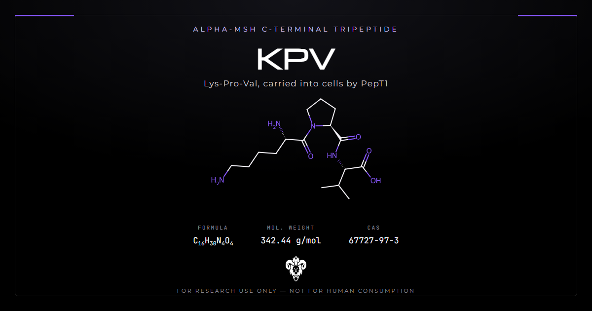
KPV is the last three residues of alpha-melanocyte-stimulating hormone, and the field named it accordingly: alpha-MSH(11-13). The name implies a small melanocortin agonist.
Then investigators ran the tests an agonist has to pass. A melanocortin receptor antagonist failed to block it. It produced no rise in cyclic AMP. It worked in mice whose MC1 receptor does not function [1]. Meanwhile a separate group showed the tripeptide riding into cells on PepT1 [2]. PepT1 moves di- and tripeptides across membranes. It does not care what they signal.
That is the interesting fact about this compound, and most write-ups skip it. Read the receptor evidence first.
Chemical identity
Three residues, and the smallest peptide in this catalogue by some distance.
| Property | Value |
|---|---|
| Sequence | Lys-Pro-Val |
| Common names | KPV, alpha-MSH(11-13), lysine-proline-valine |
| Molecular formula | C16H30N4O4 |
| Molecular weight | 342.43 |
| CAS | 67727-97-3 |
| PubChem CID | 125672 |
| InChIKey | YSPZCHGIWAQVKQ-AVGNSLFASA-N |
| Physical form | White lyophilised powder |
| Parent peptide | alpha-MSH, 13 residues, from proopiomelanocortin |
Three residues is small enough to matter
Most peptides in research use run to a dozen residues or more. At that length the sequence is close to unique. A tripeptide is not. Lys, Pro and Val in any order give the same formula and the same mass. That changes what verification has to do, and the point returns near the end of this article.
Where the name comes from
Proopiomelanocortin is cleaved into a family of peptides, one of which is alpha-MSH. Alpha-MSH runs 13 residues and acts on the melanocortin receptors MC1R through MC5R, raising cyclic AMP. Its anti-inflammatory reputation is long-standing and reasonably well evidenced [3].
Work in the late 1980s narrowed that anti-inflammatory activity toward the carboxy terminus. Investigators kept truncating and the activity kept surviving, down to the final three residues. So the field acquired a tripeptide named for its position in a hormone. The naming carried an assumption: that a fragment of a receptor agonist agonises the same receptor.
The assumption was reasonable and it did not hold
Two decades of melanocortin pharmacology have since separated the core sequence from the C-terminal one. The core of alpha-MSH, His-Phe-Arg-Trp, behaves like a melanocortin ligand. KPV does not, and the tests that show it are specific.
The receptor tests it fails
The strongest single paper here dissected the core and C-terminal peptides side by side in crystal-induced peritonitis [1]. Four results point the same way.
| Test | What an MC agonist should do | What KPV did |
|---|---|---|
| MC3/MC4 antagonist SHU9119 | Effect blocked | Effect not blocked |
| Cyclic AMP in macrophages | Rises | No increase |
| Macrophage KC and IL-1beta release | Inhibited | Not inhibited |
| Recessive yellow (e/e) mice, nonfunctional MC1R | Effect lost | Effect retained |
| Selective MC1R agonist MS05, same model | Comparable effect | MS05 gave no reduction |
Alpha-MSH and the MC3/4 agonist MTII behaved as melanocortin ligands throughout that study. KPV reduced leukocyte accumulation and did none of the receptor-linked things. The authors concluded the tripeptide is unlikely to work through those receptors.
The gut work repeated the key control
A German group tested KPV in two colitis models, DSS and CD45RB-high transfer [4]. They included mice carrying a nonfunctional MC1 receptor. Treated animals recovered earlier and regained weight. Inflammatory infiltrates fell, and so did myeloperoxidase activity in colonic tissue. The MC1R-deficient animals responded too. Different laboratory, different disease model, same answer.
The keratinocyte work found no second messenger
A Sheffield group measured cyclic AMP in HaCaT cells and in normal human keratinocytes [5]. They tested alpha-MSH, KPV, KP-D-V and ACTH. None of the peptides raised it. Intracellular calcium did respond across a wide concentration range, but only with an adenosine agonist present to suppress the cyclic AMP pathway. Then they transfected MC1R into CHO cells, and a calcium response to both alpha-MSH and the KPV peptides appeared. The receptor can couple to these peptides. Keratinocytes were not doing it.
What it is instead: a PepT1 substrate
PepT1 is an intestinal transporter for di- and tripeptides. It sits at high levels in the small intestine and at low levels in the healthy colon. Inflammatory bowel disease induces it in the colon.
An Emory group ran the transport experiments directly [2]. They used tritiated KPV to establish uptake kinetics. Unlabelled KPV then competed against a known PepT1 substrate. Nanomolar concentrations suppressed NF-kappaB and MAP kinase signalling in intestinal epithelial cells and in Jurkat T cells. Proinflammatory cytokine output fell with it. Given in drinking water, it reduced inflammation in both DSS and TNBS colitis.
Why the route changes the reading
A receptor ligand acts at the cell surface and the dose that matters is the concentration outside. A transported substrate has to be carried in, and the dose that matters is whatever the transporter delivers. That makes transporter expression a variable in every KPV experiment. Expression here is not constant. The disease under treatment raises it.
The nanomolar figure is the part worth noticing
Signalling effects at nanomolar concentrations are unusual for a molecule this small with no identified receptor. The antibacterial data needs micromolar concentrations, a thousandfold higher. That gap marks a different phenomenon rather than a stronger version of the same one.
The transporter is not a neutral courier
Here the story acquires a complication that the promotional literature never mentions.
A Georgia State group asked what PepT1 itself does in colitis-associated cancer [6]. They used mice overexpressing human PepT1 in intestinal epithelium, and mice with the transporter deleted. Overexpressing animals developed larger tumours, a greater tumour burden and more intestinal inflammation. Deleted animals developed fewer and smaller tumours with less inflammation. Proliferating crypt cells tracked the same way.
The same paper reported therapeutic benefit from KPV in that model. Both findings come from one group and one set of experiments. Still, more of the transporter meant more disease in that system, and KPV depends on that transporter. Nobody has reconciled the two observations. Hold them together rather than quoting whichever suits.
Most modern KPV papers test a carrier, not the peptide
Search the recent literature and the titles look like a run of positive results. Read the methods and a pattern appears: the variable under test is usually a delivery system.
| Study | What was actually the variable |
|---|---|
| Hyaluronic acid nanoparticles, 2017 [7] | Particle formulation, ~272 nm, targeting colonic epithelium and macrophages |
| Double-network hydrogel, 2022 [8] | A gel that adheres to inflamed rather than healthy mucosa; KPV described as a model drug |
| Self-immolative conjugates, 2026 [9] | A chemistry for surviving the gastrointestinal tract during oral dosing |
In each case the comparison carrying the paper is carrier-plus-peptide against free peptide. Free KPV is the weaker arm. That is the intended result. The authors set out to improve delivery and they succeeded.
What that means for reading a title
“KPV alleviates ulcerative colitis” in a paper from the last decade often means the carrier did the work and the tripeptide was the cargo. The finding is real, and it is a finding about formulation. One implication is worth stating plainly. These authors judged the free peptide insufficient on its own, several times over, and built something to fix that.
The antibacterial activity is a separate mechanism
A Delhi group compared fragments of alpha-MSH against Staphylococcus aureus [10]. The C-terminal fragments, alpha-MSH(6-13) and alpha-MSH(11-13), killed more than 90% of both methicillin-sensitive and methicillin-resistant cells at micromolar concentrations. At nanomolar they killed around half. The N-terminal fragment alpha-MSH(1-5) did nothing. Activity survived sodium chloride and divalent cations, which defeat many antimicrobial peptides. The killing mechanism was membrane depolarisation and permeabilisation.
Membrane disruption has nothing to do with NF-kappaB suppression. Two activities, two mechanisms, two concentration ranges. Merging them is the most common error in secondary write-ups of this compound. The antibacterial route puts KPV in the company of membrane-active peptides like LL-37, not of signalling molecules.
Outside the gut
The colitis literature dominates, and the other models are smaller and more scattered.
Traumatic brain injury
A Mainz group gave a single intraperitoneal dose of 1 mg/kg thirty minutes after controlled cortical impact in mice [11]. Lesion volume came out smaller. The careful part is what did not change. TNF-alpha and IL-1beta expression held steady, and so did the total count of Iba-1 positive cells. Only the branching of those microglia changed, indicating an altered activation state rather than fewer cells. That study also tracked MC1R expression, which rose threefold by 12 hours after injury.
Corneal epithelium
Topical KPV accelerated corneal epithelial wound healing in rabbits after mechanical abrasion, with nitric oxide implicated in the effect [12]. Wound healing in the eye is a different endpoint from inflammation suppression. Nobody has extended this line of work much since.
The D-proline analogue is a different compound
KP-D-V, sometimes written KDPV, substitutes D-proline for L-proline. A Dundee group studied it in fetal alveolar epithelial cells [13]. After endotoxin, it suppressed nuclear localisation of the NF-kappaB subunits p50, p65 and p75. They proposed antagonism at the interleukin-1 receptor as the route. Note the framing. An IL-1 receptor antagonist, and a stereoisomer of the compound this article is about.
What the studies actually used
Amounts are worth collecting in one place, because they span four orders of magnitude and three routes.
| Study | Species and model | Route and amount |
|---|---|---|
| Dalmasso 2008 [2] | Mouse, DSS and TNBS colitis | Drinking water; nanomolar in the cell work |
| Schaible 2013 [11] | Mouse, controlled cortical impact | Single intraperitoneal dose, 1 mg/kg, 30 min after injury |
| Bonfiglio 2006 [12] | Rabbit, corneal abrasion | Topical, 1, 5 or 10 mg/ml, 30 microlitres, four times daily for 4 days |
| Singh 2011 [10] | S. aureus, in vitro | Micromolar for more than 90% kill; nanomolar for around half |
| Xiao 2017 [7] | Mouse, ulcerative colitis | Oral, loaded into 272 nm hyaluronic acid nanoparticles |
Two things stand out. The signalling work and the bactericidal work sit a thousandfold apart. And the in vivo entries use different routes in different species, so none of them cross-validates another.
Several key papers do not state a dose in the abstract
The peritonitis dissection [1] and the two colitis models in the German study [4] report the design and the outcome without an amount in the abstract. Anyone building a picture of exposure needs the full texts, and the numbers above are what the abstracts support.
Two experiments that would settle the interpretation
Neither of these is a recommendation. They are the gaps a reader should notice.
Give KPV to a PepT1-knockout animal with colitis
PepT1-knockout mice exist and one group has already used them, but for a question about cancer rather than about this peptide [6]. Dosing them and measuring colonic inflammation would test the transport hypothesis directly. If the anti-inflammatory effect survives without the transporter, the uptake story is incomplete. If it disappears, the mechanism is settled. That control has not been published.
Match the delivered concentration, then compare carrier against free peptide
In the formulation literature the free peptide always loses. That comparison is not clean, because the carrier changes how much peptide reaches the tissue and how long it stays there. Matching the delivered concentration and then comparing would separate two claims that currently travel together: that the carrier improves delivery, and that the carrier improves the response for a given amount delivered. Only the first has been demonstrated.
How to read a KPV study
Four questions separate a study that can attribute a mechanism from one that can only report an outcome.
Did it test the receptor?
Dosing KPV and measuring inflammation establishes nothing about mechanism. A study using MC1R-deficient mice, an antagonist such as SHU9119, or a cyclic AMP readout can attribute. A study using PepT1 knockouts or transport competition can attribute on the other side. Everything else is an outcome measurement.
Was the peptide the variable, or the carrier?
Check whether free KPV appeared as its own arm and how it performed. In the formulation literature it usually appears and usually underperforms, which is the point of those papers.
What route and what compartment?
Oral in drinking water, intraperitoneal, intracolonic and topical are different experiments. The transporter argument only applies where PepT1 is expressed. A topical corneal result and an oral colitis result do not support each other.
What concentration?
Nanomolar signalling effects and micromolar bactericidal effects are separate regimes. A claim that borrows evidence across that gap is borrowing across mechanisms too.
Verifying research material
Identity confirmation for KPV works differently from a longer peptide, and the reason is arithmetic.
Mass does not identify a tripeptide
Lys-Pro-Val, Lys-Val-Pro, Pro-Lys-Val, Pro-Val-Lys, Val-Lys-Pro and Val-Pro-Lys all share the formula C16H30N4O4 and the mass 342.43. So does KP-D-V, which differs only in the stereochemistry at proline. A mass spectrum consistent with 342.43 fits at least seven distinct compounds. One of them has its own separate literature.
Sequencing, or a retention-time comparison against a characterised reference on the same method, is what distinguishes them. This is the check that matters for KPV and it is the one most often skipped.
Free acid or amide
Much of the older literature uses capped analogues: KPV-NH2 and Ac-KPV-NH2 appear throughout the conformational and structure-activity work. The C-terminal amide shifts the mass by one unit against the free acid. It also changes the charge state at the carboxy terminus. Material sold as KPV is normally the free acid. Confirm which form a given paper used before mapping its results onto it.
Handling
The lyophilised solid is hygroscopic. Equilibrate it to room temperature before opening, which keeps condensation out of the vial. Proline-containing peptides hold up well. With no cysteine and no methionine, KPV has no oxidation route of the kind that dominates larger peptides. Reconstituted solutions still degrade faster than the solid. Every batch we supply carries a certificate of analysis recording the identity and purity data behind it.
Common questions about KPV
Identity and origin
Is KPV a melanocortin agonist? The published receptor tests say no. It is a fragment of a melanocortin agonist, which is a statement about sequence rather than pharmacology [1][4].
Where does the sequence come from? Residues 11 to 13 of alpha-MSH, which is itself cleaved from proopiomelanocortin.
What is the relationship to GHK-Cu? None beyond both being short peptides studied for tissue effects. GHK-Cu is a copper-binding tripeptide with an entirely separate literature and mechanism.
Evidence
Which finding is best supported? Reduction of colonic inflammation in rodent models. More than one group has replicated it, across DSS, TNBS and transfer colitis [2][4].
What is the strongest caution? PepT1, the transporter it depends on, promoted tumour development when overexpressed in a colitis-associated cancer model [6].
Is there human evidence? No controlled human trials of KPV are indexed. The compound sits at the preclinical stage across every indication discussed here.
Handling and verification
Does purity by HPLC settle identity? No. A tripeptide’s sequence isomers coelute poorly or well depending on the method, and they share a mass. Purity and identity are separate questions here.
What should a COA show? Formula, mass, and something that fixes the sequence rather than only the composition.
Does the free acid or the amide matter for comparison? Yes. The two forms differ by a single mass unit and by the charge at the C-terminus. Papers using capped analogues are studying a related compound.
Is a melanocortin receptor assay useful here? Only as a negative control. The published receptor work found no coupling in the cells where the anti-inflammatory effect appears [1][5].
Summary of the evidence
KPV is a tripeptide named for its position in alpha-MSH. Its activity survived a truncation series. Its mechanism did not follow the parent. Across two laboratories and three disease models, it works in animals lacking a functional MC1 receptor, resists blockade by a melanocortin antagonist, and raises no cyclic AMP. What it does do is enter cells through PepT1, a transporter the inflamed colon expresses more of. Inside, it suppresses NF-kappaB and MAP kinase signalling at nanomolar concentrations.
Two qualifications belong with that. PepT1 is not a passive courier: more of it made colitis-associated cancer worse in the model where it was tested. And most of the recent literature is delivery research. There the free peptide is the control arm rather than the subject.
Kimera Chems supplies KPV alongside related peptides including BPC-157, with full analytical documentation. More compounds in this class are covered in our peptides research library.
Research use only. Not for human or veterinary use. Nothing here describes a therapy or a dosing protocol.
References
- Getting SJ, Schiöth HB, Perretti M. Dissection of the anti-inflammatory effect of the core and C-terminal (KPV) alpha-melanocyte-stimulating hormone peptides. J Pharmacol Exp Ther. 2003;306(2):631-7. PMID 12750433. DOI
- Dalmasso G, Charrier-Hisamuddin L, Nguyen HT, Yan Y, Sitaraman S, Merlin D. PepT1-mediated tripeptide KPV uptake reduces intestinal inflammation. Gastroenterology. 2008;134(1):166-78. PMID 18061177. DOI
- Brzoska T, Luger TA, Maaser C, Abels C, Böhm M. Alpha-melanocyte-stimulating hormone and related tripeptides: biochemistry, antiinflammatory and protective effects in vitro and in vivo, and future perspectives for the treatment of immune-mediated inflammatory diseases. Endocr Rev. 2008;29(5):581-602. PMID 18612139. DOI
- Kannengiesser K, Maaser C, Heidemann J, Luegering A, Ross M, Brzoska T, Bohm M, Luger TA, et al. Melanocortin-derived tripeptide KPV has anti-inflammatory potential in murine models of inflammatory bowel disease. Inflamm Bowel Dis. 2008;14(3):324-31. PMID 18092346. DOI
- Elliott RJ, Szabo M, Wagner MJ, Kemp EH, MacNeil S, Haycock JW. alpha-Melanocyte-stimulating hormone, MSH 11-13 KPV and adrenocorticotropic hormone signalling in human keratinocyte cells. J Invest Dermatol. 2004;122(4):1010-9. PMID 15102092. DOI
- Viennois E, Ingersoll SA, Ayyadurai S, Zhao Y, Wang L, Zhang M, Han MK, Garg P, et al. Critical role of PepT1 in promoting colitis-associated cancer and therapeutic benefits of the anti-inflammatory PepT1-mediated tripeptide KPV in a murine model. Cell Mol Gastroenterol Hepatol. 2016;2(3):340-357. PMID 27458604. DOI
- Xiao B, Xu Z, Viennois E, Zhang Y, Zhang Z, Zhang M, Han MK, Kang Y, et al. Orally Targeted Delivery of Tripeptide KPV via Hyaluronic Acid-Functionalized Nanoparticles Efficiently Alleviates Ulcerative Colitis. Mol Ther. 2017;25(7):1628-1640. PMID 28143741. DOI
- Zhao Y, Xue P, Lin G, Tong M, Yang J, Zhang Y, Ran K, Zhuge D, et al. A KPV-binding double-network hydrogel restores gut mucosal barrier in an inflamed colon. Acta Biomater. 2022;143:233-252. PMID 35245681. DOI
- Cheng J, Wu P, Li C, Han Y, Sun M, Dou Y, Chen S, Zhang J. Inflammation-triggered self-immolative conjugates enable oral peptide delivery by overcoming gastrointestinal barriers. Sci Adv. 2026;12(3):eaea2989. PMID 41533788. DOI
- Singh M, Mukhopadhyay K. C-terminal amino acids of alpha-melanocyte-stimulating hormone are requisite for its antibacterial activity against Staphylococcus aureus. Antimicrob Agents Chemother. 2011;55(5):1920-9. PMID 21282427. DOI
- Schaible EV, Steinsträßer A, Jahn-Eimermacher A, Luh C, Sebastiani A, Kornes F, Pieter D, Schäfer MK, et al. Single administration of tripeptide α-MSH(11-13) attenuates brain damage by reduced inflammation and apoptosis after experimental traumatic brain injury in mice. PLoS One. 2013;8(8):e71056. PMID 23940690. DOI
- Bonfiglio V, Camillieri G, Avitabile T, Leggio GM, Drago F. Effects of the COOH-terminal tripeptide alpha-MSH(11-13) on corneal epithelial wound healing: role of nitric oxide. Exp Eye Res. 2006;83(6):1366-72. PMID 16965771. DOI
- Haddad JJ, Lauterbach R, Saadé NE, Safieh-Garabedian B, Land SC. Alpha-melanocyte-related tripeptide, Lys-d-Pro-Val, ameliorates endotoxin-induced nuclear factor kappaB translocation and activation: evidence for involvement of an interleukin-1beta193-195 receptor antagonism in the alveolar epithelium. Biochem J. 2001;355(Pt 1):29-38. PMID 11256945. DOI
Research use only. Not for human or veterinary use. Nothing here describes a therapy or a dosing protocol.