
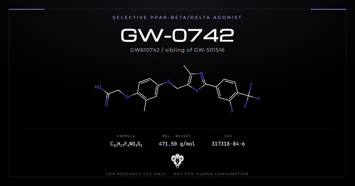
GW-0742 reaches an EC50 of 1.1 nanomolar at PPAR-delta with roughly thousandfold selectivity over the other two receptor subtypes [1]. On paper that makes it one of the cleanest nuclear receptor tools available.
Then the control experiments arrive. Its vasorelaxant effect survives in tissue from mice that lack the receptor [2]. Its aortic effects in one diabetic rat model run through a non-genomic route [3]. Its protection in experimental colitis appeared in animals given no ligand at all [4].
None of that makes the compound useless. It makes it a compound that has to be used with the controls attached, which is the most interesting thing about it as research material. Kimera supplies GW-0742 as a reference standard for that kind of work.
Everything below reports findings from cell and animal models. This material is for research use only, not for human or veterinary use.
What GW-0742 is
The molecule is a thiazole-linked phenoxyacetic acid. Its carboxylic acid head binds the receptor’s ligand pocket, and the fluorinated aryl thiazole tail carries the selectivity.
Identity and physical data
| Property | Value |
|---|---|
| Compound | GW-0742 |
| Synonyms | GW610742, GW 0742X |
| PubChem CID | 9934458 |
| CAS number | 317318-84-6 |
| Molecular formula | C21H17F4NO3S2 |
| Molecular weight | 471.5 g/mol |
| InChIKey | HWVNEWGKWRGSRK-UHFFFAOYSA-N |
| IUPAC name | 2-[4-[[2-[3-fluoro-4-(trifluoromethyl)phenyl]-4-methyl-1,3-thiazol-5-yl]methylsulfanyl]-2-methylphenoxy]acetic acid |
| Chemical class | Phenoxyacetic acid, thiazole |
| Stereochemistry | None; the InChIKey stereo block is UHFFFAOYSA |
Where the potency figure comes from
The 2003 paper that introduced this series screened lipophilic carboxylic acids, then optimised the hits by structure-guided design [1]. Two compounds came out of that work. Compound 7k became GW-501516 and compound 7l became GW-0742, and the paper reports the 1.1 nanomolar figure with thousandfold subtype selectivity for the latter.
The sibling compound
GW-501516 came from the same optimisation round and reached the wider literature first. The two differ in the aryl substitution on the thiazole. Anyone comparing published potencies should check which compound a paper used, since the names are close enough to blur in a citation chain.
The receptor it was built for
PPAR-beta/delta is a nuclear hormone receptor that heterodimerises with RXR and regulates fatty acid oxidation, lipid handling and inflammatory gene expression. It is expressed almost everywhere, which is part of why attributing an effect to it is hard.
The canonical target genes
Two transcripts anchor most of this work: angiopoietin-like 4 and adipose differentiation-related protein, written Angptl4 and Adrp [5]. Both rise in wild-type mouse colon after treatment and stay flat in receptor-null colon, and chromatin immunoprecipitation ties the change to receptor occupancy at those promoters [5].
Why that matters for experimental design
Angptl4 and Adrp give a positive control that reports receptor engagement directly. Measuring them alongside a phenotype tells you whether the compound reached its target in that tissue. Skipping them leaves a phenotype with no engagement evidence behind it.
Expression is the confound
Wide expression cuts both ways. It explains why an agonist produces effects in almost any tissue somebody tests, and it removes the simplest attribution argument available for other receptors: that the tissue does not carry the target.
Nothing about a positive result in a new organ is surprising here. The receptor is present in that organ, and so is a long list of other proteins a lipophilic carboxylic acid can touch (PubMed). Surprise is not the test. Controls are.
Attribution controls, and why this compound needs them
The antagonist tool
GSK3787 is an irreversible PPAR-beta/delta antagonist characterised in 2010 [5]. Given orally, it blocked GW-0742-driven Angptl4 and Adrp induction in wild-type colon. A second antagonist, GSK0660, appears in the cardiac literature [6]. Both give a pharmacological way to test whether an effect runs through the receptor.
The null-mouse standard
Genetic deletion is the stronger test. The PPAR-beta/delta-null mouse has carried most of the definitive work in this field, and several results below exist only because someone ran the compound in both genotypes (PubMed).
What a complete control set looks like
Three elements. A target gene readout to prove engagement, an antagonist arm to test pharmacological dependence, and a null-genotype arm to test genetic dependence. A paper reporting a GW-0742 phenotype with none of the three has shown you an effect of the molecule, not an effect of the receptor.
Where the attribution failed
Vasorelaxation in receptor-null tissue
A 2010 study found the compound relaxed both systemic and pulmonary vessels, and limited right heart hypertrophy in rats with hypoxia-induced pulmonary hypertension [2]. The authors then tested tissue from genetically modified mice. The dilator effect persisted without the receptor, and without cell surface prostacyclin receptors [2]. That is the clearest published example of the problem.
A non-genomic route in diabetic rat aorta
A 2018 study worked on aortic rings from streptozotocin-treated rats and measured contraction and dilation in organ baths [3]. Nuclear receptors act through transcription, which takes hours. Effects on vascular tone in an organ bath appear in minutes. The timescale alone rules the receptor out, and the authors describe the effects as non-genomic and off-target [3].
Colitis protection without a ligand
A 2007 study induced colitis with dextran sodium sulfate in wild-type and receptor-null mice, with and without the compound [4]. Protection tracked receptor genotype rather than ligand treatment. The title states the conclusion plainly: the mechanism is ligand-independent [4]. Receptor expression mattered; adding an agonist did not add much.
How to read those three together
They fail in different directions. One phenotype is receptor-independent, one is transcription-independent, and one is ligand-independent. Each failure implicates a different assumption, which is why a single control arm is not enough for this compound.
Findings in metabolic models
Incretin production
A 2011 study in Gastroenterology examined proglucagon regulation in murine GLUTag and human NCI-H716 L cells, then in wild-type and receptor-null mice [7]. Receptor activation raised GLP-1 production in the cell models. The in vivo genotype comparison is what makes this one of the better-controlled results in the set.
Diet, exercise and the mimetic label
A 2019 study combined aerobic exercise and agonist treatment in diet-induced obese female mice and measured immunometabolic endpoints [8]. The paper calls agonists of this receptor exercise mimetics, a label this class has carried for a decade. Read it as shorthand for overlapping transcriptional programmes in rodents. It is not a claim about people, and the study measured mice.
Fatty acid oxidation is the through-line
Read the positive results across organs and one process recurs. Cardiac protection ran through fatty acid oxidation-linked respiration [10]. Astrocyte work measured beta-oxidation directly [17]. The receptor’s established transcriptional programme covers lipid handling, so a metabolic readout is the one most likely to reflect genuine receptor engagement.
That gives a practical rule for designing an experiment with GW-0742. Choose an endpoint the receptor is known to control, then confirm engagement with a target gene. An endpoint far from lipid metabolism needs a stronger control set, not a weaker one.
Endothelial metabolism depends on context
A 2020 study compared the compound against VEGF-A in endothelial cells and found the metabolic response depended on the context the cells sat in [9]. Two stimuli, one cell type, divergent outcomes. Context dependence of that kind is a warning against pooling endothelial results across preparations.
Findings in cardiac and mitochondrial models
Ischaemia and reperfusion in rat heart
A 2021 study treated rats with the agonist, the antagonist GSK0660, or both, then subjected hearts to global ischaemia and reperfusion ex vivo [6]. Protection came with ALDH2 upregulation, less oxidative stress and preserved mitochondrial energy production. The antagonist arm is present in this design, which puts it above much of the surrounding literature.
Respiratory function
A 2022 follow-up traced the same protection to fatty acid oxidation-linked respiration and PGC-1-alpha with NRF-1 signalling [10]. Mitochondrial respirometry gives a quantitative endpoint rather than a histological one, and the mechanism it points at is the receptor’s established transcriptional programme.
Diabetic cardiomyopathy
A 2024 study reported reduced inflammation and fibrosis in a diabetic cardiomyopathy model [11]. Fibrosis endpoints take weeks to develop, so this branch of the work sits at the opposite end of the timescale from the organ bath results above.
Findings in tumour and hepatic models
Several papers examine whether receptor activation changes tumour development in rodents. Each result belongs to its model and its carcinogen.
Skin and colon
A 2008 study reported inhibition of chemically induced skin tumorigenesis after ligand activation [12]. A related paper found the receptor and COX2 inhibition attenuated colon carcinogenesis through independent signalling mechanisms [13]. Independent means the two arms did not converge, which is itself an attribution result.
Cell proliferation in keratinocytes
A 2008 study in human HaCaT keratinocytes reported inhibited proliferation after ligand activation [14]. Cell line work of this kind sets the concentration range that later animal work uses.
Liver
A 2016 study reported suppressed liver tumorigenesis in hepatitis B transgenic mice [15]. An earlier study found attenuated carbon tetrachloride hepatotoxicity with downregulated proinflammatory gene expression [16]. Both are model-bound observations in rodents.
What the tumour work does and does not say
Chemical carcinogenesis models answer a narrow question. They ask whether a treatment changes tumour incidence or multiplicity after a defined initiator, in one strain, in one tissue. They do not rank a compound against anything used clinically, and they do not transfer between tissues.
The value here is comparative. Skin, colon and liver were tested by overlapping groups using shared reagents [12][13][15]. Reading them together shows which findings reproduce across tissue and which do not, and that is a more useful question than whether any single result was positive.
Neural, glial and inflammatory work
A 2019 study measured fatty acid oxidation in astrocytes carrying the PSEN1 delta-E9 mutation and reported improvement after treatment [17]. Astrocyte metabolism is a plausible receptor target, since these cells run substantial beta-oxidation.
A 2025 study compared agonists across PPAR subtypes in dextran sodium sulfate colitis and tracked gut microbiota alongside the inflammatory endpoints [18]. Comparing subtypes in one experiment is the design that separates a receptor effect from a class effect.
Reading a GW-0742 paper in five minutes
The literature runs past 200 records and the quality varies. Four questions sort it quickly.
Which controls ran
Look for a genotype arm, an antagonist arm, or a target gene measurement. One of the three is adequate for a preliminary result. None of the three means the paper reports what the molecule did, not what the receptor did.
What concentration, and for how long
Note the concentration against the 1.1 nanomolar EC50 [1]. Note the exposure time too. A response inside ten minutes cannot be transcriptional, which is the reasoning that identified the aortic results as non-genomic [3].
Which compound the authors actually used
GW-0742 and GW-501516 are adjacent in name and in structure [1]. Citation chains blur them. Check the methods section rather than the abstract.
Whether the endpoint suits the receptor
Lipid handling, fatty acid oxidation and inflammatory gene expression sit inside the receptor’s known programme. Vascular tone and cell proliferation sit further out, and those are the areas where attribution has failed [2][3][14].
Physicochemical properties and handling
| Property | Detail |
|---|---|
| Appearance | White to off-white crystalline solid |
| Solubility | Soluble in DMSO and ethanol; poorly soluble in water |
| Recommended stock | DMSO, split into single-use aliquots |
| Storage, solid | Controlled room temperature, sealed and desiccated |
| Storage, solution | Prepare fresh in single-use aliquots rather than holding stock |
| Stability note | Carboxylic acid; keep dry and avoid strong base |
| Handling | Standard laboratory controls for a fine powder |
Storage guidance is a house recommendation. Analytical documentation is per-lot release testing.
The carboxylic acid changes the solution chemistry
A free acid ionises with pH. Solubility in aqueous buffer rises above the pKa and falls below it, so a stock that looks fine at pH 7.4 can precipitate on dilution into an acidic medium. Check for precipitate under the microscope before blaming a null result on the compound, and match vehicle concentration across every well.
Concentration ranges in the literature
Cell work in this literature clusters in the high nanomolar to low micromolar band, well above the 1.1 nanomolar EC50 [1]. Anything approaching 10 micromolar leaves the selective range, and the off-target results discussed above were seen at higher concentrations. Report the concentration used and treat potency and selectivity as separate questions.
Checks that catch a bad stock
Three cheap ones. Inspect for precipitate after dilution into the working medium. Run a target gene readout on a known-responsive cell line before committing to an experiment. And confirm the solid dissolved fully before the first aliquot, since an incompletely dissolved stock makes every downstream concentration wrong in the same direction.
Silent failure is the risk worth guarding against. A poorly dissolved carboxylic acid gives a stock that looks correct, reads correct on the label, and shifts every downstream concentration in the same direction. Nothing in the resulting data flags it.
Analytical characterization
Mass and ultraviolet detection
Electrospray mass spectrometry in negative mode gives a deprotonated ion at m/z 470.1 for the C21H17F4NO3S2 formula, and the acid ionises well that way. The aryl thiazole chromophore supports routine HPLC purity work with ultraviolet detection. Batch documentation for catalogue material sits in the certificate of analysis database.
Fluorine as a confirmation channel
Four fluorines sit on this molecule: a trifluoromethyl group and a ring fluorine. Fluorine-19 NMR gives a clean, uncluttered spectrum for that arrangement, and it is a faster identity confirmation than a full proton assignment. Few catalogue compounds offer that channel, and this one does.
What an impurity profile should separate
The thioether linkage is the synthetic joint. Its two halves, the thiazolylmethyl fragment and the substituted phenoxyacetic acid, are the likely process impurities, and both differ enough in polarity to resolve under reversed-phase conditions. The free acid and any residual ester also separate cleanly.
Where GW-0742 sits among nuclear receptor tools
| Compound | Receptor target | Class |
|---|---|---|
| GW-0742 | PPAR-beta/delta agonist | Phenoxyacetic acid, thiazole |
| GW-501516 | PPAR-beta/delta agonist | Phenoxyacetic acid, thiazole |
| SR-9009 | REV-ERB agonist | Pyrrole carboxamide |
| SR-9011 | REV-ERB agonist | Pyrrole carboxamide |
All four appear in the same transcriptional literature and two of them share a scaffold. Further reading sits in the metabolic research library.
What this literature does not establish
No human data
Published work on this compound is preclinical. No controlled human trial results are indexed.
Receptor attribution is unsettled per phenotype
Attribution has to be argued phenotype by phenotype. Some results carry genotype controls [4][5][7]. Others explicitly fail them [2][3]. Treating the whole literature as one body of receptor pharmacology is the mistake this compound invites, and reading each paper’s control set is the way around it.
Publication bias sits on top of everything
Positive results dominate this record, as they dominate most agonist literatures. The failed attributions reached print because the authors went looking for them, and a study that finds nothing rarely gets written up at all. Treat the balance of published results as evidence about what people measured, not about how often the receptor is responsible.
Concentration reporting is uneven
Papers differ in how precisely they state exposure, and the gap between the nanomolar EC50 and the micromolar working concentrations is wide. Two studies reporting the same phenotype at different concentrations may be describing different mechanisms.
The class label carries assumptions
The exercise mimetic framing came from rodent transcriptional overlap [8]. It is a convenient label and it has never been a description of a human outcome.
Frequently asked questions
How selective is GW-0742?
The introducing paper reports an EC50 of 1.1 nanomolar at PPAR-delta with roughly thousandfold selectivity over the other subtypes [1].
How does it differ from GW-501516?
Both came out of the same 2003 optimisation series and differ in aryl substitution on the thiazole [1]. Check which one a paper used before comparing numbers.
Which genes confirm the compound reached its target?
Angptl4 and Adrp. Both rise with receptor engagement and stay flat in receptor-null tissue [5].
Why do some published effects survive receptor deletion?
Because they were never receptor effects. Vasorelaxation persisted in null tissue [2], and aortic responses in one model act on a non-genomic timescale [3].
What antagonists pair with it?
GSK3787, characterised as an irreversible antagonist [5], and GSK0660, used in the cardiac ischaemia work [6].
Is GW-0742 an approved drug anywhere?
No. It is a preclinical research compound with no clinical approval, and PubMed indexes no controlled human trial results for it.
Which target genes should a positive control measure?
Angptl4 and Adrp, both established as receptor-dependent in wild-type against null tissue [5]. Measuring one of them costs a qPCR plate and turns a phenotype into an attributed result.
What is the practical difference from a REV-ERB agonist?
Different receptor family and different mechanism. PPAR-beta/delta is a ligand-activated transcription factor that heterodimerises with RXR; REV-ERB agonists act on a repressor arm of the circadian machinery.
How should stocks be prepared?
DMSO, single-use aliquots, prepared fresh rather than held. Watch for precipitation when diluting the free acid into acidic aqueous media.
References
- Sznaidman ML, Haffner CD, Maloney PR, et al. Novel selective small molecule agonists for peroxisome proliferator-activated receptor delta (PPARdelta): synthesis and biological activity. Bioorg Med Chem Lett. 2003;13(9):1517-21. PubMed DOI
- Harrington LS, Moreno L, Reed A, et al. The PPARbeta/delta agonist GW0742 relaxes pulmonary vessels and limits right heart hypertrophy in rats with hypoxia-induced pulmonary hypertension. PLoS One. 2010;5(3):e9526. PubMed DOI
- Perez-Diaz N, Pushkarsky I, Oweis N, et al. The non-genomic effects of the PPAR beta/delta agonist GW0742 on streptozotocin treated rat aorta. Curr Mol Pharmacol. 2018;11(2):149-154. PubMed DOI
- Hollingshead HE, Morimura K, Adachi M, et al. PPARbeta/delta protects against experimental colitis through a ligand-independent mechanism. Dig Dis Sci. 2007;52(11):2912-9. PubMed DOI
- Palkar PS, Borland MG, Naruhn S, et al. Cellular and pharmacological selectivity of the peroxisome proliferator-activated receptor-beta/delta antagonist GSK3787. Mol Pharmacol. 2010;78(3):419-30. PubMed DOI
- Papatheodorou I, Galatou E, Panagiotidis GD, et al. Cardioprotective effects of PPARbeta/delta activation against ischemia/reperfusion injury in rat heart are associated with ALDH2 upregulation, amelioration of oxidative stress and preservation of mitochondrial energy production. Int J Mol Sci. 2021;22(12):6399. PubMed DOI
- Daoudi M, Hennuyer N, Borland MG, et al. PPARbeta/delta activation induces enteroendocrine L cell GLP-1 production. Gastroenterology. 2011;140(5):1564-74. PubMed DOI
- Le Garf S, Murdaca J, Mothe-Satney I, et al. Complementary immunometabolic effects of exercise and PPARbeta/delta agonist in the context of diet-induced weight loss in obese female mice. Int J Mol Sci. 2019;20(20):5182. PubMed DOI
- Faulkner A, Lynam E, Purcell R, et al. Context-dependent regulation of endothelial cell metabolism: differential effects of the PPARbeta/delta agonist GW0742 and VEGF-A. Sci Rep. 2020;10(1):7849. PubMed DOI
- Papatheodorou I, Makrecka-Kuka M, Kuka J, et al. Pharmacological activation of PPARbeta/delta preserves mitochondrial respiratory function in ischemia/reperfusion via stimulation of fatty acid oxidation-linked respiration and PGC-1alpha/NRF-1 signaling. Front Endocrinol (Lausanne). 2022;13:941822. PubMed DOI
- Rostami A, Palomer X, Pizarro-Delgado J, et al. PPARbeta/delta prevents inflammation and fibrosis during diabetic cardiomyopathy. Pharmacol Res. 2024;210:107515. PubMed DOI
- Bility MT, Devlin-Durante MK, Blazanin N, et al. Ligand activation of peroxisome proliferator-activated receptor beta/delta inhibits chemically induced skin tumorigenesis. Carcinogenesis. 2008;29(12):2406-14. PubMed DOI
- Hollingshead HE, Borland MG, Billin AN, et al. Ligand activation of peroxisome proliferator-activated receptor-beta/delta and inhibition of cyclooxygenase 2 attenuate colon carcinogenesis through independent signaling mechanisms. Carcinogenesis. 2008;29(1):169-76. PubMed DOI
- Borland MG, Foreman JE, Girroir EE, et al. Ligand activation of peroxisome proliferator-activated receptor-beta/delta inhibits cell proliferation in human HaCaT keratinocytes. Mol Pharmacol. 2008;74(5):1429-42. PubMed DOI
- Balandaram G, Kramer LR, Kang BH, et al. Ligand activation of peroxisome proliferator-activated receptor-beta/delta suppresses liver tumorigenesis in hepatitis B transgenic mice. Toxicology. 2016;363-364:1-9. PubMed DOI
- Shan W, Palkar PS, Murray IA, et al. Ligand activation of peroxisome proliferator-activated receptor beta/delta attenuates carbon tetrachloride hepatotoxicity by downregulating proinflammatory gene expression. Toxicol Sci. 2008;105(2):418-28. PubMed DOI
- Konttinen H, Gureviciene I, Oksanen M, et al. PPARbeta/delta-agonist GW0742 ameliorates dysfunction in fatty acid oxidation in PSEN1 delta-E9 astrocytes. Glia. 2019;67(1):146-159. PubMed DOI
- Li JH, Xu J, Hu JX, et al. PPARgamma/beta/delta agonists can ameliorate dextran sodium sulfate-induced colitis and modulate gut microbiota. J Gastroenterol Hepatol. 2025;40(6):1536-1547. PubMed DOI