
The analogue is not a stronger version of the hormone. At the type-1 receptor it binds slightly worse than native IGF-1. The people who made it said so in the paper that introduced it [1].
Its extra potency in culture comes from somewhere else. Native IGF-1 spends almost all of its time captured by binding proteins, and IGF-1 LR3 escapes that capture. Same signal, brake removed.
The name misleads in a second way. “Long” describes a 13-residue extension on the front of the molecule, not a duration of action. One study has tracked the analogue in a living animal by mass spectrometry. IGF-1 LR3 vanished from rat serum faster than its shorter relatives [19].
Chemical identity
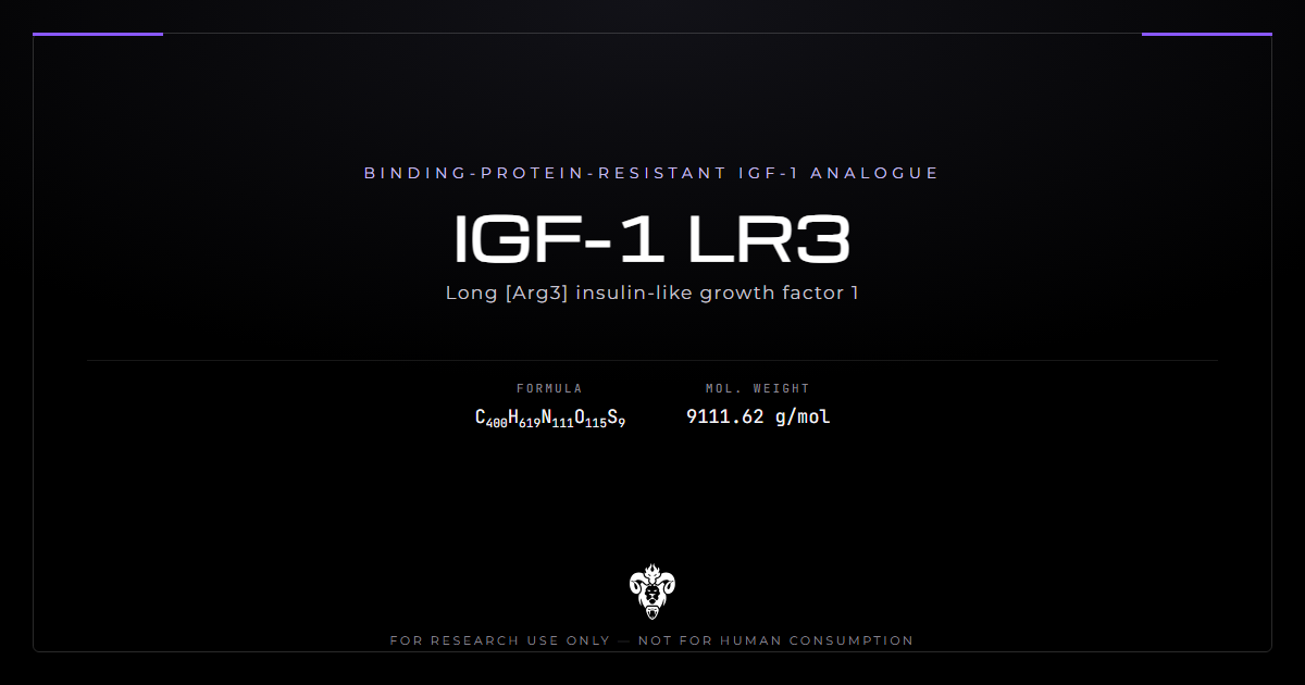
An 83-residue recombinant protein: human IGF-1 with one substitution and an N-terminal extension borrowed from pig growth hormone.
| Property | Value |
|---|---|
| Full name | Long [Arg3] insulin-like growth factor 1 |
| Common names | IGF-1 LR3, LongR3 IGF-I, LR3 IGF-1 |
| Residues | 83 |
| Molecular formula | C400H619N111O115S9 |
| Average mass | 9,111.6 Da |
| Disulfide bonds | 3 |
| Parent protein | Human IGF-1, 70 residues, 7,648.8 Da |
| Parent entry | UniProt P05019 residues 49-118 |
| Substitution | Glu3 to Arg |
| Extension | Met-Phe-Pro-Ala-Met-Pro-Leu-Ser-Ser-Leu-Phe-Val-Asn |
| Extension origin | Methionyl porcine growth hormone, residues 1-11, plus Val-Asn |
| Expression host | Escherichia coli |
The two modifications
Both changes serve the same purpose, and neither touches the receptor-binding surface directly.
Glu at position 3 becomes Arg. That single swap collapses affinity for the binding proteins. King and colleagues found the substituted analogues binding bovine IGFBP-2 very poorly [1].
The 13-residue extension came from the expression system rather than from a design goal. Francis and colleagues fused the first 11 residues of methionyl porcine growth hormone plus Val-Asn to the front [2]. They reported that the hydrophobic extension helped the protein fold correctly at high yield. It also lowers binding protein affinity further.
What the name does not mean
Three misreadings of the label are worth clearing up before the evidence.
“Long” is the fusion extension. It does not describe half-life, and no half-life advantage over IGF-1 has been demonstrated in an animal by direct measurement.
“LR3” is Long plus Arg3. A related analogue, R3-IGF-I, carries the substitution without the extension, and Des(1-3)-IGF-I removes the first three residues instead. All three appear in the same literature and behave differently [19].
The molecule is human IGF-1 in its receptor-binding core. Anything the native hormone does through the type-1 receptor, IGF-1 LR3 also does. That is why the analogue works as a reagent, and why its risks track the pathway rather than the modification.
The binding protein system it evades
Six high-affinity binding proteins control how much IGF reaches a receptor. That system is the target of the design, so it is worth understanding before the results.
The ternary complex
Most circulating IGF-1 is not free. It sits in a 150 kDa complex with IGFBP-3 and an acid-labile subunit. That complex is what keeps a potent mitogen in the blood without constant signalling.
Higaki and colleagues showed what happens when the complex thins out [10]. In streptozotocin-diabetic rats with reduced IGFBP-3, injected IGF-1 disappeared faster than in normal rats at every dose tested.
The binding proteins also govern where the hormone goes. Perfusing isolated beating rat hearts with IGF-1 LR3 cut uptake of labelled IGF-1 to 9%. IGFBP-3 binding sites accounted for a further share of transport [15].
Why cells make more binding protein when you feed them IGF
The system is a loop, not a fixed buffer, and that is the part the analogue defeats.
Grimes and Hammond gave ovarian granulosa cells IGF-1 and watched them respond by producing more IGFBP-3 and IGFBP-2 [3]. Feeding the pathway increases the buffer that restrains it.
IGF-1 LR3 was significantly more potent than IGF-1 at driving that response [3]. The analogue is not exempt from the feedback. It triggers the same loop and then ignores the output.
What the design actually achieved
Two 1992 papers from the same Adelaide group define the compound. Together they make an argument that is easy to state backwards.
Receptor affinity went down
King and colleagues compared the analogues against native IGF-1 on rat L6 myoblasts (PMID 1311930). Both Gly3 and Arg3 versions bound the type-1 receptor slightly less well than IGF-1 itself.
Their conclusion is explicit. Reduced binding to the binding proteins, rather than increased receptor binding, explains the greater biological potency of the analogues [1].
Potency went up anyway
Francis and colleagues then tested the fusion versions across cell types [2]. In L6 myoblasts, every analogue beat native IGF-1 at stimulating protein and DNA synthesis and at inhibiting protein breakdown.
The ordering carries the argument. Potency ranked highest in cell lines that secrete binding proteins into the medium [2]. That is exactly where escaping capture would help most.
Independent work reproduced the pattern in other systems. Rat ovarian theca-interstitial cells showed greater DNA synthesis with IGF-1 LR3 and Des(1-3)-IGF-I than with IGF-1 [8]. Xi and colleagues later ran the reverse experiment in L6 myogenic cells. Neutralising IGFBP-3 raised IGF-1-stimulated proliferation by 58%, and IGF-1 LR3-stimulated proliferation by only 33% [17].
That asymmetry is the mechanism restated. Removing the buffer helps the molecule that was still subject to it.
Where the analogue does not win
The potency advantage is conditional, and three studies show what happens when the condition is absent.
Zhao and colleagues compared IGF-1, Des(1-3)-IGF-I and the Long Arg3 analogue on bovine immune cells (PMID 7508487). At 12.5 µg/L, hydrogen peroxide release from neutrophils rose 65% with IGF-1 and 64% with Des(1-3). The analogue gave 32% [5].
The mononuclear cell result went further. IGF-1 and Des(1-3)-IGF-I both stimulated thymidine incorporation at 100 µg/L, and the Long Arg3 analogue did not [5].
Neff and colleagues found the same equivalence in a developmental model. Chick embryos were treated during the period of normal motoneuron death. IGF-1, IGF-II, Des(1-3)-IGF-I and the analogue all rescued motoneurons to a comparable degree [6].
Retinal cultures gave the third example. IGF-1 and three analogues with differing receptor and binding protein affinities all inhibited neuroretinal cell death at 50 ng/mL. The authors read that as evidence the protective effect depends on neither affinity [14].
Take those together with the myoblast data. IGF-1 LR3 wins where binding proteins are abundant in the medium. It draws or loses where they are not, which is what the design predicts and what the marketing omits.
Built for a flask
The analogue’s largest real-world application is a cell culture reagent. The published record reflects that far more than any physiological use.
Serum-free CHO culture
Insulin is the standard growth factor for serum-free Chinese hamster ovary cultures, and it has limits.
Morris and Schmid compared insulin against the analogue in two recombinant CHO lines expressing cytokine receptors [13]. One line grew without any added growth factor, the other required insulin or the analogue.
Under production conditions the analogue sustained viability better than insulin in both lines [13]. The authors named it the preferred growth factor for those cells. That is a bioprocessing conclusion rather than a biological one.
Stem cell and differentiation media
The same substitution logic applies wherever binding proteins in conditioned medium would otherwise soak up the added factor.
Bieberich reported replacing insulin with the analogue to differentiate embryonic stem cells into neuroprogenitors and insulin-secreting cells [16]. Media formulation is the setting, and the analogue’s value there is stability of effect rather than potency as such.
Note what this means for the literature as a whole. A large share of published work uses IGF-1 LR3 as a tool for interrogating the binding protein system. The analogue is the instrument, not the subject.
In vivo rodent findings
Animal work exists, and it is more specific and less flattering than the reagent literature might suggest.
| Study | Model | Route | Finding |
|---|---|---|---|
| Harel 1992 [4] | Adult male rats | Intracerebroventricular | No change in pulsatile GH secretion |
| Conlon 1995 [7] | Female guinea pigs | 7-day infusion | Organ weights up, body weight unchanged |
| Staley 1998 [11] | Newborn rat pups | Dietary | Villus height and cell counts increased |
| Levolger 2019 [18] | C26 tumour-bearing mice | Systemic | Muscle preserved, tumour growth accelerated |
| Mongongu 2021 [19] | Rats | Intramuscular, 100 µg/kg | Undetectable after 4 hours |
Organ-selective, not general growth
Conlon and colleagues infused guinea pigs for seven days with recombinant IGF-1, IGF-II or the analogue [7].
Body weight gain, feed intake, feed conversion and carcass composition were unaffected by any treatment. The analogue did raise the fractional weights of adrenals, gut, kidneys and spleen. Overall growth still did not move [7].
A separate result points the same way. Adult rats received the analogue centrally, at several doses. The pulsatile pattern of growth hormone release did not change [4].
Gut and intestine
The intestinal effect is the most reproducible one in the rodent record.
Staley and colleagues reared newborn rat pups artificially and fed them the analogue [11]. Villus height and cell counts rose above milk-replacer controls, and above what the replacer alone produced.
That result belongs to a specific window. It describes a neonatal gut adapting to an oral growth factor. Nothing in it transfers to an adult animal or another tissue.
Muscle, and what it cost
One study measured muscle directly under a systemic challenge, and it reported both halves of the outcome.
Levolger and colleagues ran a C26 cancer cachexia model in mice and included the analogue as a treatment arm [18]. It limited loss of muscle mass. The paper states the trade-off plainly: that came at the expense of accelerated tumour growth.
Both observations belong to tumour-bearing mice. Neither transfers to any other setting. The authors’ preferred intervention in that model was an activin-receptor kinase inhibitor rather than the growth factor.
What the antidoping literature found
Sport authorities prohibit IGF-1 and its analogues, and that has produced the only modern analytical work on the compound.
Detection windows
Mongongu and colleagues validated an immunopurification and high-resolution mass spectrometry method, then dosed rats intramuscularly at 100 µg/kg (PMID 33587816).
Des(1-3)-IGF-I and R3-IGF-I remained detectable to 24 hours. IGF-1 LR3 disappeared rapidly after 4 hours [19].
The fragments outlasted the parent. A previously undescribed degradation product, Des(1)-LongR3-IGF-I, appeared alongside Des(1-10) and Des(1-11) forms. The last of those stayed detectable to 16 hours [19].
Read that against the name. The N-terminal extension that defines the analogue is also the part proteases remove first. In this experiment the intact molecule was the shortest-lived of the three.
Product quality in seized material
The same paper examined black market products directly, and the finding shaped its method.
Abundant signs of lower quality and oxidised peptide forms were present [19]. The authors concluded that any assay has to monitor both native and mono-oxidised forms to work on real-world samples.
Methionine oxidation is the obvious suspect. The extension contributes two methionine residues to a protein that otherwise carries one. So the modification that defines the analogue triples its most oxidation-prone residue count.
The brake it removes exists for a reason
Binding proteins are not packaging. They carry their own biology, and some of it runs against the growth signal.
Nickerson and colleagues showed IGFBP-3 inducing apoptosis in MCF7 breast cancer cells. IGF-1 LR3 blocked that apoptosis more potently than IGF-1 did [9]. The analogue’s defining property is precisely what makes it the better blocker.
Butt and colleagues reviewed the wider picture [12]. They describe type-1 receptor signalling as a potent survival function, and IGFBP-3 as carrying pro-apoptotic effects of its own. Kerr and Baxter restate the same architecture in current terms [20]. The binding proteins govern bioavailability and also influence cancer progression through receptor-independent routes.
Nothing there is a claim about any person. It is the reason a reagent that defeats bioavailability control is a laboratory tool and not a therapeutic.
What has never been done
Several gaps are worth naming precisely, because their absence is often mistaken for reassurance.
No human pharmacokinetic study describes this analogue. Absorption, clearance and half-life in people are unmeasured, and the antidoping work is a rat experiment [19].
No controlled human trial exists at all. The compound was never approved for human use anywhere, which the antidoping literature states directly [19].
No head-to-head animal comparison establishes a duration advantage over IGF-1. The one direct measurement points the other way [19].
Structure-activity coverage is also thin. The two 1992 papers remain the primary characterisation [1][2]. Since then the analogue has been used as an instrument rather than examined as a subject.
How to read an IGF-1 LR3 study
Four questions decide whether a result means what it appears to mean.
Which analogue?
Long-R3, R3 and Des(1-3) are three different molecules with different behaviour in the same assay [19]. Papers abbreviate inconsistently, so check the methods rather than the abstract.
Does the system contain binding proteins?
The analogue’s advantage appears only where binding proteins are present to be escaped [2]. In a system with none, it should perform like IGF-1 or slightly worse. In retinal cultures every analogue tested behaved alike [14].
Reagent or subject?
Much of the literature uses IGF-1 LR3 to remove a variable. In the heart perfusion work it served to block IGF-1 binding sites [15]. Those papers report on the system, not on the analogue.
In vitro or in vivo?
Culture potency and animal outcome diverge sharply here. Higher potency in a flask [2][8] sits alongside organ-selective effects and no overall growth in a guinea pig [7].
Verifying research material
A disulfide-bonded 83-residue protein expressed in bacteria carries a specific set of failure modes, and the certificate should address them.
Identity
Intact mass is the first check. The analogue runs at 9,111.6 Da against 7,648.8 Da for mature IGF-1. That 1,463 Da difference is one no instrument can miss.
Peptide mapping resolves the rest. The Arg3 substitution changes a tryptic fragment. The substituted arginine introduces a cleavage site the glutamate version lacks.
Oxidation deserves separate attention. Seized material carried oxidised forms in the antidoping study [19]. Each oxidised methionine adds 16 Da, which appears as a satellite peak beside the parent.
Purity and folding
Three disulfide bonds mean correct folding is a purity question rather than a formality.
Reversed-phase chromatography separates misfolded disulfide isomers from correctly folded material, since they share a mass and differ in hydrophobicity. A certificate reporting only SDS-PAGE cannot distinguish them.
Bacterial expression brings its own asks. Host cell protein, residual endotoxin and incomplete cleavage of the fusion partner all belong on the certificate [2].
Handling
Lyophilised protein should be stored cold, dry and dark, and reconstituted immediately before use.
Adsorption to plastic is the routine loss at working concentrations, which for this analogue are nanomolar. Aliquot on reconstitution rather than freezing and thawing a single vial repeatedly.
Carrier protein matters more here than for small peptides. Dilute solutions of a 9 kDa protein in plain buffer lose material to surfaces. The binding proteins that would normally stabilise the native hormone are the very thing this analogue avoids.
Kimera publishes third-party certificates of analysis for every lot in its COA database. Laboratories source IGF-1 LR3 as a binding-protein-resistant reference for IGF axis work. It appears alongside Ipamorelin and CJC-1295, where the growth hormone axis is the shared theme. It also pairs with Selank as a contrasting short peptide. Related work appears in the peptides category.
Common questions about IGF-1 LR3
What is the difference from IGF-1? An Arg for Glu swap at position 3 and a 13-residue N-terminal extension from porcine growth hormone [1][2].
Is it more potent at the receptor? No. It binds the type-1 receptor slightly less well than IGF-1, and its potency comes from escaping the binding proteins [1].
Does the name mean it lasts longer? “Long” refers to the extension. In rats it became undetectable after 4 hours, faster than R3 and Des(1-3) forms [19].
What was it designed for? Cell culture. It replaces insulin in serum-free CHO and stem cell media [13][16].
What do animal studies show? Organ-selective effects without overall growth in guinea pigs [7]. Intestinal growth in newborn rat pups [11]. Muscle preservation with accelerated tumour growth in a mouse cachexia model [18].
Is there human data? None. No controlled trial and no pharmacokinetic study exist for this analogue [19].
Summary of the evidence
Identity: 83 residues, C400H619N111O115S9, 9,111.6 Da, three disulfides. Human IGF-1 with Glu3 to Arg and a 13-residue porcine growth hormone extension [1][2].
Mechanism: reduced binding protein affinity, not increased receptor affinity. The originators state it explicitly [1].
Culture potency: higher than IGF-1 in cell lines that secrete binding proteins [2], and comparable where none are present [14].
Feedback: cells respond to IGF stimulation by making more binding protein. The analogue drives that response harder and then ignores it [3].
Intended use: a serum-free culture reagent, preferred over insulin for sustaining CHO viability under production conditions [13].
Animal record: no overall growth in guinea pigs despite raised organ weights [7], and no change in pulsatile growth hormone release [4]. Neonatal intestinal growth [11], and muscle preservation alongside accelerated tumour growth in tumour-bearing mice [18].
Duration: undetectable in rat serum after 4 hours, with N-terminally clipped fragments outlasting the intact molecule [19].
Limits: no human data of any kind, and thin structure-activity coverage since 1992. Oxidised low-quality material has been documented in seized products [19].
Status: supplied for laboratory research use only.
References
- King R, Wells JR, Krieg P, Snoswell M, Brazier J, Bagley CJ, Wallace JC, Ballard FJ, Ross M, Francis GL. Production and characterization of recombinant insulin-like growth factor-I (IGF-I) and potent analogues of IGF-I, with Gly or Arg substituted for Glu3, following their expression in Escherichia coli as fusion proteins. J Mol Endocrinol. 1992;8(1):29-41. PMID 1311930. DOI
- Francis GL, Ross M, Ballard FJ, Milner SJ, Senn C, McNeil KA, Wallace JC, King R, Wells JR. Novel recombinant fusion protein analogues of insulin-like growth factor (IGF)-I indicate the relative importance of IGF-binding protein and receptor binding for enhanced biological potency. J Mol Endocrinol. 1992;8(3):213-223. PMID 1378742. DOI
- Grimes RW, Hammond JM. Insulin and insulin-like growth factors (IGFs) stimulate production of IGF-binding proteins by ovarian granulosa cells. Endocrinology. 1992;131(2):553-558. PMID 1379161. DOI
- Harel Z, Tannenbaum GS. Synergistic interaction between insulin-like growth factors-I and -II in central regulation of pulsatile growth hormone secretion. Endocrinology. 1992;131(2):758-764. PMID 1639021. DOI
- Zhao X, McBride BW, Trouten-Radford LM, Burton JH. Effects of insulin-like growth factor-I and its analogues on bovine hydrogen peroxide release by neutrophils and blastogenesis by mononuclear cells. J Endocrinol. 1993;139(2):259-265. PMID 7508487. DOI
- Neff NT, Prevette D, Houenou LJ, Lewis ME, Glicksman MA, Yin QW, Oppenheim RW. Insulin-like growth factors: putative muscle-derived trophic agents that promote motoneuron survival. J Neurobiol. 1993;24(12):1578-1588. PMID 8301266. DOI
- Conlon MA, Tomas FM, Owens PC, Wallace JC, Howarth GS, Ballard FJ. Long R3 insulin-like growth factor-I (IGF-I) infusion stimulates organ growth but reduces plasma IGF-I, IGF-II and IGF binding protein concentrations in the guinea pig. J Endocrinol. 1995;146(2):247-253. PMID 7561636. DOI
- Duleba AJ, Spaczynski RZ, Olive DL, Behrman HR. Effects of insulin and insulin-like growth factors on proliferation of rat ovarian theca-interstitial cells. Biol Reprod. 1997;56(4):891-897. PMID 9096870. DOI
- Nickerson T, Huynh H, Pollak M. Insulin-like growth factor binding protein-3 induces apoptosis in MCF7 breast cancer cells. Biochem Biophys Res Commun. 1997;237(3):690-693. PMID 9299428. DOI
- Higaki K, Matsumoto Y, Fujimoto R, Kurosaki Y, Kimura T. Pharmacokinetics of recombinant human insulin-like growth factor-I in diabetic rats. Drug Metab Dispos. 1997;25(11):1324-1327. PMID 9351911.
- Staley MD, Gibson CA, Herbein JF, Grosvenor CE, Baumrucker CR. Rat milk and dietary long arginine3 insulin-like growth factor I promote intestinal growth of newborn rat pups. Pediatr Res. 1998;44(4):512-518. PMID 9773839. DOI
- Butt AJ, Firth SM, Baxter RC. The IGF axis and programmed cell death. Immunol Cell Biol. 1999;77(3):256-262. PMID 10361258. DOI
- Morris AE, Schmid J. Effects of insulin and LongR3 on serum-free Chinese hamster ovary cell cultures expressing two recombinant proteins. Biotechnol Prog. 2000;16(5):693-697. PMID 11027158. DOI
- Seigel GM, Chiu L, Paxhia A. Inhibition of neuroretinal cell death by insulin-like growth factor-1 and its analogs. Mol Vis. 2000;6:157-163. PMID 10973501.
- Boes M, Dake BL, Booth BA, Sandra A, Bateman M, Knudtson KL, Bar RS. IGF-I and IGFBP-3 transport in the rat heart. Am J Physiol Endocrinol Metab. 2003;284(1):E237-E239. PMID 12485812. DOI
- Bieberich E. Replacement of insulin by LongR3-IGF-1 allows for the differentiation of ES cells into neuroprogenitors and insulin-secreting cells. Anal Biochem. 2005;346(1):185-187. PMID 16169509. DOI
- Xi G, Kamanga-Sollo E, Hathaway MR, Dayton WR, White ME. Effect of constitutive expression of porcine IGFBP-3 on proliferation and differentiation of L6 myogenic cells. Domest Anim Endocrinol. 2006;31(1):35-51. PMID 16233971. DOI
- Levolger S, Wiemer EAC, van Vugt JLA, Huisman SA, van Vledder MG, van Damme-van Engel S, Ambagtsheer G, IJzermans JNM, de Bruin RWF. Inhibition of activin-like kinase 4/5 attenuates cancer cachexia associated muscle wasting. Sci Rep. 2019;9(1):9826. PMID 31285507. DOI
- Mongongu C, Coudoré F, Domergue V, Ericsson M, Buisson C, Marchand A. Detection of LongR3-IGF-I, Des(1-3)-IGF-I, and R3-IGF-I using immunopurification and high resolution mass spectrometry for antidoping purposes. Drug Test Anal. 2021;13(7):1256-1269. PMID 33587816. DOI
- Kerr A, Baxter RC. Noncoding RNA actions through IGFs and IGF binding proteins in cancer. Oncogene. 2022;41(25):3385-3393. PMID 35597813. DOI
IGF-1 LR3 is sold for laboratory research use only. Not for human consumption, nor medical, veterinary, or household uses.
Literature retrieved from PubMed.