
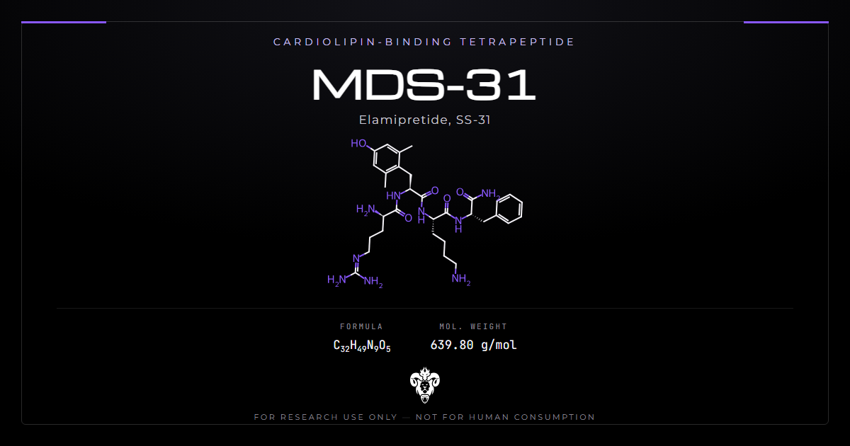
MDS-31 is a four-residue peptide with an unusually crowded set of names. Identifying it correctly changes what its literature is worth.
The sequence is D-Arg-Dmt-Lys-Phe-NH2. The published record calls that molecule SS-31, elamipretide, MTP-131 and Bendavia, depending on the decade and the author. MDS-31 is not an obscure research chemical. It reached named clinical trials under an international nonproprietary name [14].
That matters because the literature is substantial and specific. MDS-31 has a defined molecular target that is not a protein. It has a mechanism worked out at the level of membrane biophysics, and a preclinical record spanning kidney, eye, heart and vasculature.
What follows covers the identity and its aliases, the target, the mechanism in detail, the structure-activity work, the preclinical and clinical records, and how to verify the material.
Chemical identity: what you are actually handling
MDS-31 is a tetrapeptide with two non-standard features.
| Property | Value |
|---|---|
| Sequence | D-Arg-Dmt-Lys-Phe-NH2 |
| INN | Elamipretide |
| Aliases | SS-31, MTP-131, Bendavia, MDS-31 |
| Molecular formula | C32H49N9O5 |
| Molecular weight | 639.79 g/mol |
| PubChem CID | 11764719 |
| InChIKey | SFVLTCAESLKEHH-WKAQUBQDSA-N |
| Residue 1 | D-arginine, inverted stereochemistry |
| Residue 2 | 2,6-dimethyltyrosine, non-proteinogenic |
| C-terminus | Primary amide, not free acid |
| Molecular target | Cardiolipin, a phospholipid |
The aliases are not interchangeable in search
Anyone researching MDS-31 needs all four names. SS-31 dominates the mechanistic literature and elamipretide the clinical. MTP-131 appears in the ophthalmic work [6], and Bendavia in older cardiovascular material.
A search on the catalogue code alone returns almost nothing. A search on SS-31 returns the bulk of the mechanism papers. Missing that connection is the commonest reason MDS-31 gets described as understudied.
Three modifications, three purposes
The peptide carries three departures from an ordinary tetrapeptide, and each does specific work.
D-arginine at position 1 inverts the stereochemistry of the N-terminal residue. Peptidases recognise L-amino acids, so a D-residue at the terminus blocks aminopeptidase attack.
The C-terminal primary amide removes the free carboxylate, which similarly blocks carboxypeptidase attack and removes a negative charge.
2,6-dimethyltyrosine is the most interesting substitution. The two methyl groups flanking the phenol restrict rotation and increase hydrophobicity. This residue supplies the aromatic bulk that drives membrane insertion.
The overall architecture alternates cationic and aromatic residues, which is the defining feature of the Szeto-Schiller peptide class [12].
What MDS-31 targets: a lipid, not a protein
Most peptide therapeutics bind receptors. This one binds a phospholipid, and that difference shapes everything about how it behaves.
Cardiolipin
Cardiolipin is an anionic phospholipid expressed almost exclusively on the inner mitochondrial membrane. It carries four acyl chains rather than the usual two. That unusual geometry lets it stabilise the curved cristae membranes where oxidative phosphorylation happens [3].
Cardiolipin also organises the respiratory complexes into supercomplexes, improving electron transfer efficiency [3]. Reports of cardiolipin peroxidation and depletion span a range of conditions associated with energy deficiency [3].
Cardiolipin sits in one membrane of one organelle. A compound binding it selectively therefore acquires organelle targeting without needing a targeting sequence.
The cytochrome c switch
The mechanistic core of this compound concerns a single protein doing two different jobs.
Cytochrome c normally shuttles electrons within the respiratory chain. Forming a hydrophobic complex with cardiolipin switches it from electron carrier to peroxidase [2]. That peroxidase activity catalyses cardiolipin peroxidation, damaging the membrane. It also impedes electron flux and inhibits ATP synthesis [2].
The switch is doubly damaging. It destroys the lipid organising the cristae, and it removes a carrier from the electron transport chain.
It is also self-reinforcing, which is the feature that makes it worth targeting. Peroxidised cardiolipin binds cytochrome c differently, promoting further conversion to the peroxidase form. Damage therefore accelerates rather than plateauing, and an intervention that interrupts the cycle early prevents more than an intervention applied late.
That kinetic asymmetry explains a recurring pattern in the animal work below. Pretreatment protocols produce larger effects than treatment applied after injury is established.
Mechanism
Birk and colleagues established the binding using a polarity-sensitive fluorescent analogue of the peptide [1].
MDS-31 binds cardiolipin with high affinity through electrostatic and hydrophobic interactions [3]. Nuclear magnetic resonance showed the aromatic residues penetrating deep into cardiolipin-containing bilayers, at roughly a 1:1 ratio [2].
Selective inhibition without loss of function
The pharmacologically elegant part is what the peptide does to the cytochrome c complex.
It inhibits the peroxidase activity while protecting the electron carrying function [2]. Both activities belong to the same protein, so suppressing one without the other is a non-obvious result.
The protective effect works partly by shielding the heme iron [1]. Functionally, MDS-31 restored cytochrome c reduction and mitochondrial oxygen consumption in the presence of added cardiolipin. In fresh mitochondria it increased state 3 respiration and the efficiency of ATP synthesis [2].
The electrostatic account
Later biophysical work refined the picture and moved it away from a simple binding model.
Mitchell and colleagues analysed interactions with model and mitochondrial membranes [10]. MDS-31 partitions into the membrane interfacial region, with affinity and binding density related directly to surface charge.
Two findings shaped the revised mechanism. Binding did not destabilise lamellar bilayers even at the highest concentrations, though it did alter lipid packing saturably [10]. And the peptide modulated surface electrostatics in both model and mitochondrial membranes [10].
The authors proposed that tuning surface charge underpins the protective properties. Altering the distribution of ions and basic proteins at the membrane interface is the suggested route [10]. As proof of concept, the peptide shifted divalent calcium distribution in that region and reduced the energetic burden of calcium stress in mitochondria [10].
That reframing matters for experimental design. A mechanism operating through bulk membrane electrostatics behaves differently from one using a discrete binding site. Concentration dependence differs most of all.
Structure-activity relationships
One study directly tested which structural features matter, and its results argue against treating the peptide class as interchangeable.
Mitchell and colleagues compared three tetrapeptide analogues against SS-31, varying aromatic side chain composition and sequence register [12]. They also produced the first structural models for the class, using nuclear magnetic resonance and molecular dynamics.
The structural result was unexpected. All analogues except SS-31 formed compact reverse turn conformations in the membrane-bound state [12]. SS-31 itself did not.
All four bound cardiolipin-containing membranes. They differed significantly in equilibrium binding behaviour and membrane interactions, and most notably in their effects on membrane surface charge [12].
Function tracked those differences imperfectly. None of the peptides had strict requirements for side chain composition or sequence register in order to permeate cells and reach mitochondria. They did differ significantly in their ability to restore mitochondrial membrane potential, preserve ATP content and promote cell survival [12]. The tryptophan-containing analogue had the strongest effect on membrane properties and the greatest efficacy in cell culture [12].
Read practically, that means cell penetration and mitochondrial targeting hold up across the class. Functional efficacy does not.
How the compound was found
The discovery route explains why the mechanism took so long to pin down.
Szeto and Birk describe the finding as serendipitous [4]. The compounds emerged from work on a different problem, and their selective accumulation in mitochondria was observed before anyone knew what they bound.
That order of events left a decade in which MDS-31 had demonstrable effects and no agreed mechanism. The cardiolipin account arrived later, and the electrostatic refinement later still.
Szeto frames the resulting class as one that restores mitochondrial plasticity, meaning the capacity of the electron transport chain to raise ATP output when metabolic demand rises or fuel supply falls [4]. That capacity declines in many age-associated conditions [9].
The framing matters for interpretation. A compound restoring reserve capacity should show little effect in a healthy system operating well within its limits, and larger effects under stress. Reviews of the class make exactly that claim, describing effects in disease models without corresponding effects in normal healthy organisms [4][9].
The preclinical record
The animal literature is broad, and the organ range follows from the target: any tissue dense in mitochondria is a candidate.
Kidney
Ischaemia causes acute kidney injury through ATP depletion. Recovery depends on how fast ATP returns on reperfusion [1].
Pretreatment protected cristae membranes during renal ischaemia and prevented mitochondrial swelling [1]. Prompt ATP recovery allowed rapid repair of ATP-dependent processes, including restoration of the actin cytoskeleton and cell polarity. It also inhibited apoptosis and protected tubular barrier function [1].
A companion study addressed the vasculature rather than the tubules. Loss of microvascular density is implicated in progression from acute ischaemic injury to chronic kidney disease, and no established intervention exists for it [5].
Treatment prevented mitochondrial swelling and protected cristae in both endothelial and epithelial cells. It prevented no-reflow after ischaemia. At four weeks it had reduced loss of peritubular capillaries and cortical arterioles, interstitial inflammation and fibrosis [5].
Separately, mice on a high-fat diet for 28 weeks showed a similar pattern. Treatment preserved mitochondrial structure across glomerular endothelial cells, podocytes and proximal tubular epithelial cells. It restored renal AMP kinase activity, and prevented intracellular lipid accumulation, endoplasmic reticulum stress and apoptosis [7]. It had no effect on weight gain, insulin resistance or hyperglycaemia [7].
That dissociation is worth noting. The protective effect appeared without any improvement in the metabolic drivers, which places the action downstream of them.
Eye
Alam and colleagues measured spatial visual behaviour in mouse models of diabetes. They administered the compound either systemically or as eye drops [6].
Visual decline emerged before overt metabolic or ophthalmic abnormalities appeared. Treatment reversed the visual decline without improving glycaemic control or reducing body weight [6].
Again the effect appeared without correcting the upstream metabolic state.
Vasculature
In apolipoprotein E knockout mice fed a Western diet for 12 weeks, subcutaneous treatment reduced the area and size of atherosclerotic plaques. Plaque composition changed as well [8]. Markers of oxidative stress fell, superoxide dismutase activity rose, and several circulating inflammatory markers decreased [8].
Barth syndrome models
The most mechanistically specific animal work concerns a genetic disorder of cardiolipin itself.
Barth syndrome results from loss-of-function mutations in TAFAZZIN, the gene responsible for remodelling cardiolipin. The result is a dramatically increased ratio of monolysocardiolipin to mature cardiolipin [11].
In tafazzin knockdown mice, treatment improved mitochondrial respiratory capacity and promoted supercomplex organisation. The monolysocardiolipin ratio did not change [11]. The authors inferred that MDS-31 acts on respiratory chain function rather than on cardiolipin composition directly [11].
Follow-up work in the same model found the treatment restored mitochondrial morphology, affecting proteins involved in fission dynamics and mitophagy [13].
The clinical programme
This is where MDS-31 diverges from most compounds in a research catalogue.
The compound entered named clinical trials as elamipretide. A 2025 review lists PROGRESS-HF, TAZPOWER, MMPOWER-3 and ReCLAIM among them [14]. Those programmes span heart failure, Barth syndrome, primary mitochondrial myopathy and ophthalmic indications.
Two things should be said plainly about that.
Reaching phase 3 in multiple indications reflects genuine institutional confidence. It also means human pharmacokinetic and safety data exist, which most research compounds lack entirely.
It also means the compound has not been approved. A drug with completed phase 3 programmes and no marketing authorisation is one whose trials did not deliver what was needed. The review cited above summarises therapeutic potential rather than reporting trial outcomes [14]. Anyone relying on it should read the individual trial reports instead.
Kimera supplies this material for laboratory research use only, and no statement here should be read as describing clinical performance.
Physicochemical properties and handling
MDS-31 is a small, highly basic, water-soluble peptide.
Two of the four residues carry positive charge at physiological pH, arginine and lysine. The C-terminal amide removes the only negative charge an ordinary peptide would have. MDS-31 is therefore polycationic, and that charge drives its interaction with anionic membrane surfaces [10].
Aqueous solubility is high. Suppliers typically provide the peptide as a salt, commonly acetate or trifluoroacetate. Confirm which, because the counterion contributes mass and trifluoroacetate carries its own biological activity at higher concentrations.
Peptides of this size are hygroscopic. Allow vials to reach room temperature before opening, or condensation will introduce water.
Store lyophilised material sealed at low temperature. In solution the compound is more vulnerable, so prepare fresh where possible and avoid repeated freeze-thaw cycles.
Analytical characterisation
Four checks cover this peptide.
Accurate mass confirms C32H49N9O5 at 639.79. Nine nitrogens across two basic residues give a distinctive composition and a characteristic multiply-charged envelope by electrospray ionisation.
Sequence confirmation by tandem mass spectrometry. Fragmentation should show the expected ion series, and the dimethyltyrosine residue produces a mass 28 units above ordinary tyrosine.
Stereochemical verification of the D-arginine, by amino acid analysis after hydrolysis with a chiral method. An L-arginine substitution shares the molecular formula and accurate mass exactly. It would also lose the peptidase resistance the residue exists to provide.
Counterion identity and content, by ion chromatography or fluorine NMR if trifluoroacetate is suspected.
What a rigorous certificate should contain
Chromatographic purity with method conditions stated.
Accurate mass confirming the molecular formula.
Sequence confirmation by tandem mass spectrometry.
Stereochemistry at position 1, established rather than assumed.
Counterion identity and content, quantified.
Water content, given the hygroscopicity.
Kimera publishes third-party certificates of analysis for every lot in its COA database. Laboratories source MDS-31 as a cardiolipin-binding reference peptide. It sometimes sits alongside BAM15 or DADA, which act on mitochondrial bioenergetics by unrelated routes. Related chemistry appears in the peptides category.
Common misclassifications
Four errors recur.
Suppliers describe MDS-31 as an unstudied research peptide. It is elamipretide, with a mechanistic literature spanning two decades and named phase 3 trials [14].
Copy describes its target as a receptor or an enzyme. The target is cardiolipin, a phospholipid. The mechanism runs through membrane biophysics rather than receptor occupancy [10].
Sources call it an antioxidant. The mechanism is not radical scavenging. MDS-31 prevents cardiolipin from converting cytochrome c into a peroxidase, stopping peroxidation at source rather than mopping up products [2][3].
Summaries treat peptides in the class as interchangeable. Analogues differing only in aromatic side chain or sequence register showed significantly different efficacy at restoring membrane potential and promoting cell survival [12].
Experimental design considerations
Search all four names. The mechanistic literature is filed under SS-31, the clinical under elamipretide, and the catalogue code returns almost nothing.
Do not assume a discrete binding site. The evidence supports a mechanism operating through membrane surface electrostatics. Its concentration dependence differs from receptor occupancy [10].
Include a cardiolipin-deficient control where possible. A tafazzin-deficient or cardiolipin-depleted system tests target dependence directly [11][13].
Measure respiration, not just viability. The proximate effect falls on electron transport efficiency and ATP synthesis [2]. A viability endpoint alone will miss it.
Correct for counterion mass before calculating molarity.
Expect protection without correction of upstream drivers. In both the kidney and eye studies, effects appeared with no improvement in glycaemic control or body weight [6][7].
Frequently asked questions
What is MDS-31? The tetrapeptide D-Arg-Dmt-Lys-Phe-NH2, known in the literature as SS-31 and by the international nonproprietary name elamipretide. Kimera supplies it as a laboratory research material.
What does it bind? Cardiolipin, an anionic phospholipid on the inner mitochondrial membrane [1][3].
How does it work? By binding cardiolipin, it prevents the cardiolipin and cytochrome c complex from acting as a peroxidase while preserving cytochrome c as an electron carrier [2], and it modulates membrane surface electrostatics [10].
Is it an antioxidant? Not in the scavenging sense. It prevents the reaction that generates the damage rather than neutralising the products.
Has it been in human trials? Yes, under the name elamipretide, across several named programmes [14]. It holds no marketing approval.
Why the non-standard residues? D-arginine and the C-terminal amide block exopeptidase degradation. Dimethyltyrosine supplies the aromatic bulk that drives membrane insertion.
What analytical check is most often missing? Stereochemistry at the D-arginine. An L-substitution is invisible to mass spectrometry.
Why does a lipid target give organelle selectivity? Cardiolipin sits almost exclusively in the inner mitochondrial membrane. Binding it selectively therefore concentrates the peptide where that lipid is, without any targeting sequence or transporter [3].
Should effects be expected in healthy tissue? Probably not. Reviews of the class describe benefit in disease models without corresponding effects in normal healthy organisms, which fits a mechanism restoring reserve capacity rather than raising baseline output [4][9].
Summary of the evidence
Identity: D-Arg-Dmt-Lys-Phe-NH2, C32H49N9O5, 639.79 g/mol, identical to SS-31 and elamipretide.
Target: cardiolipin on the inner mitochondrial membrane, bound through electrostatic and hydrophobic interactions at roughly 1:1 [2][3].
Primary mechanism: inhibition of cytochrome c peroxidase activity with preservation of electron carrier function [2]. Modulation of membrane surface electrostatics accompanies it [10].
Functional effect: restored cytochrome c reduction and oxygen consumption, increased state 3 respiration and ATP synthesis efficiency in fresh mitochondria [2].
Preclinical breadth: renal ischaemia and microvascular protection [1][5], high-fat-diet kidney injury [7], diabetic visual decline [6], atherosclerosis [8], and Barth syndrome models [11][13].
Structure-activity: cell penetration and mitochondrial targeting tolerate side chain variation, while functional efficacy does not [12].
Clinical position: named phase 3 programmes conducted under the name elamipretide [14], with no marketing approval.
Status: supplied for laboratory research use only.
References
- Birk AV, Liu S, Soong Y, et al. The mitochondrial-targeted compound SS-31 re-energizes ischemic mitochondria by interacting with cardiolipin. J Am Soc Nephrol. 2013;24(8):1250-1261. PMID 23813215. DOI
- Birk AV, Chao WM, Bracken C, Warren JD, Szeto HH. Targeting mitochondrial cardiolipin and the cytochrome c/cardiolipin complex to promote electron transport and optimize mitochondrial ATP synthesis. Br J Pharmacol. 2014;171(8):2017-2028. PMID 24134698. DOI
- Szeto HH. First-in-class cardiolipin-protective compound as a therapeutic agent to restore mitochondrial bioenergetics. Br J Pharmacol. 2014;171(8):2029-2050. PMID 24117165. DOI
- Szeto HH, Birk AV. Serendipity and the discovery of novel compounds that restore mitochondrial plasticity. Clin Pharmacol Ther. 2014;96(6):672-683. PMID 25188726. DOI
- Liu S, Soong Y, Seshan SV, Szeto HH. Novel cardiolipin therapeutic protects endothelial mitochondria during renal ischemia and mitigates microvascular rarefaction, inflammation, and fibrosis. Am J Physiol Renal Physiol. 2014;306(9):F970-F980. PMID 24553434. DOI
- Alam NM, Mills WC, Wong AA, et al. A mitochondrial therapeutic reverses visual decline in mouse models of diabetes. Dis Model Mech. 2015;8(7):701-710. PMID 26035391. DOI
- Szeto HH, Liu S, Soong Y, et al. Protection of mitochondria prevents high-fat diet-induced glomerulopathy and proximal tubular injury. Kidney Int. 2016;90(5):997-1011. PMID 27519664. DOI
- Zhang M, Zhao H, Cai J, et al. Chronic administration of mitochondrion-targeted peptide SS-31 prevents atherosclerotic development in ApoE knockout mice fed Western diet. PLoS One. 2017;12(9):e0185688. PMID 28961281. DOI
- Szeto HH. Stealth peptides target cellular powerhouses to fight rare and common age-related diseases. Protein Pept Lett. 2018;25(12):1108-1123. PMID 30381054. DOI
- Mitchell W, Ng EA, Tamucci JD, et al. The mitochondria-targeted peptide SS-31 binds lipid bilayers and modulates surface electrostatics as a key component of its mechanism of action. J Biol Chem. 2020;295(21):7452-7469. PMID 32273339. DOI
- Russo S, De Rasmo D, Signorile A, Corcelli A, Lobasso S. Beneficial effects of SS-31 peptide on cardiac mitochondrial dysfunction in tafazzin knockdown mice. Sci Rep. 2022;12(1):19847. PMID 36400945. DOI
- Mitchell W, Tamucci JD, Ng EL, et al. Structure-activity relationships of mitochondria-targeted tetrapeptide pharmacological compounds. Elife. 2022;11:e75531. PMID 35913044. DOI
- Russo S, De Rasmo D, Rossi R, Signorile A, Lobasso S. SS-31 treatment ameliorates cardiac mitochondrial morphology and defective mitophagy in a murine model of Barth syndrome. Sci Rep. 2024;14(1):13655. PMID 38871974. DOI
- Tung C, Varzideh F, Farroni E, et al. Elamipretide: a review of its structure, mechanism of action, and therapeutic potential. Int J Mol Sci. 2025;26(3):944. PMID 39940712. DOI
MDS-31 is sold for laboratory research use only. Not for human consumption, nor medical, veterinary, or household uses.
Literature retrieved from PubMed.