
Everything below reports findings from cell cultures, isolated tissue and animals. This material is for research use only, not for human or veterinary use.
Most compounds in this catalogue need several separate explanations. Carnitine needs one.
A transporter called OCTN2 moves it out of blood and into tissue. Mutations in the gene encoding that transporter cause a rare inherited disease. Saturating it is why raising muscle carnitine is slow. And whatever the gut fails to absorb becomes food for intestinal bacteria, which is where the TMAO chemistry comes from.
Four apparently unrelated facts, one mechanism. That is the frame worth carrying through the rest of this article.
Indexed loading, tracing and performance papers exist. Those human endpoints sit outside the scope of this profile. This page does not quote their milligram figures.
Chemical identity
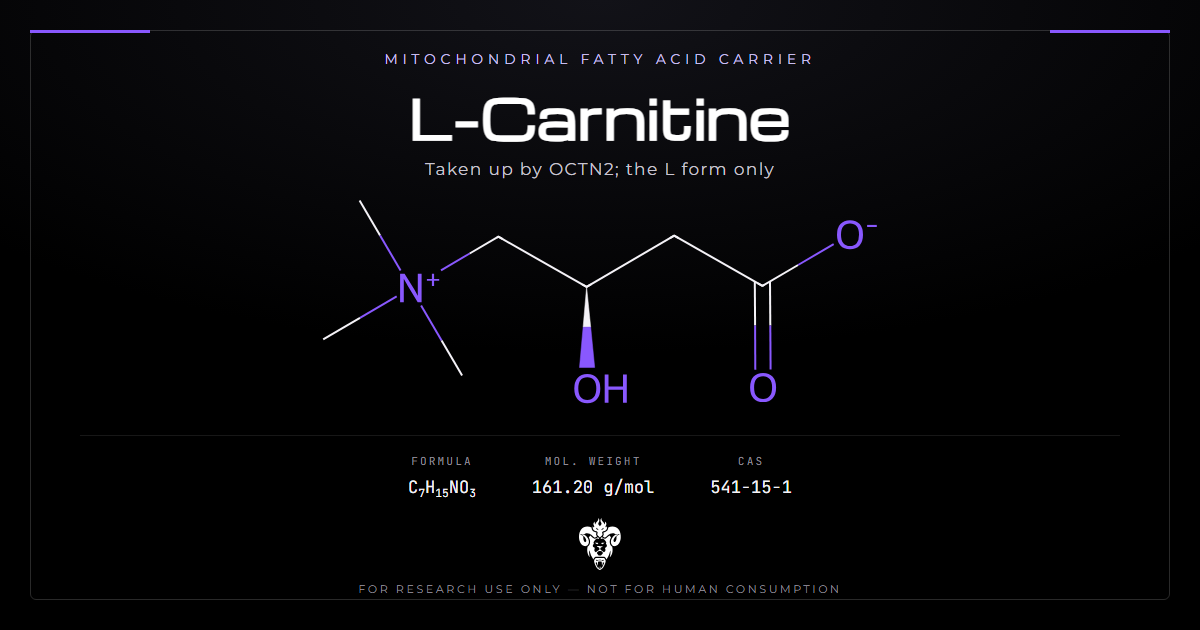
Small, charged, and water-soluble, which is the root of the delivery problem.
| Property | Value |
|---|---|
| Systematic name | (R)-3-hydroxy-4-(trimethylazaniumyl)butanoate |
| Molecular formula | C7H15NO3 |
| Molecular weight | 161.20 |
| CAS | 541-15-1 |
| PubChem CID | 10917 |
| InChIKey | PHIQHXFUZVPYII-ZCFIWIBFSA-N |
| Form | Zwitterion, quaternary ammonium and carboxylate |
| Active enantiomer | L, also written R |
The stereochemistry is not optional
That InChIKey carries a defined stereocentre. Only the L form, also written R, is biologically active. The D form is not simply inert: it competes for the same handling machinery.
So “carnitine” and “L-Carnitine” are not interchangeable terms on a label. A racemic preparation is a different material from the one every study here used.
The InChIKey for the active form ends -ZCFIWIBFSA-N. A flat key with no stereochemistry describes an unresolved or racemic material.
What carnitine actually does
Long-chain fatty acids cannot cross the inner mitochondrial membrane on their own. Carnitine carries them. An acyl group transfers onto carnitine, the acylcarnitine crosses, and the acyl group transfers back off inside.
No carnitine, no long-chain fatty acid oxidation. That is why transporter failure is a serious cellular problem rather than a subtle one.
The quaternary ammonium group is the source of everything else
A permanently charged nitrogen makes this molecule water-soluble and membrane-impermeant. It cannot diffuse into cells, so it needs a transporter. It also carries the trimethylamine group that intestinal bacteria will later strip off.
One structural feature, two consequences, and both of them run through the rest of this article.
The CPT shuttle in one page
Carnitine palmitoyltransferase I sits on the outer membrane and makes the acylcarnitine. The translocase moves that species across. CPT II on the inner face gives the acyl-CoA back and frees carnitine.
L-Carnitine is the carrier, not the enzyme. A paper that reports “carnitine raised oxidation” without naming CPT I, the translocase, or CPT II has named a substrate and skipped the machine.
Acetyl-L-carnitine and propionyl-L-carnitine already hold an acyl group. They are not the free carrier. They are different molecules. The ester section below returns to that.
Malonyl-CoA inhibits CPT I. That is how a fed cell stops long-chain oxidation. Adding free carnitine cannot override a locked CPT I. A flask that is malonyl-rich will not oxidise palmitate just because the carrier is present. Name the inhibitor state before you credit the vial.
Short-chain and medium-chain acids can skip the shuttle. They do not need this molecule. A paper that uses octanoate as the fuel and then credits carnitine has used the wrong chain length.
OCTN2 is the whole story
OCTN2, the product of the SLC22A5 gene, is the high-affinity carnitine transporter. It moves carnitine into muscle, heart, kidney and other tissue against a concentration gradient.
A functional genomics study characterised OCTN2 variants systematically and used the results to build a protein-specific variant effect predictor [1]. That is the level of attention a transporter gets when it is the only route in.
Insulin can stimulate OCTN2-dependent uptake into skeletal muscle. That fact is why indexed loading papers paired the compound with carbohydrate [4]. This page records the transporter logic. It does not reprint those protocols.
Whey protein acutely inhibited insulin-stimulated muscle uptake in a forearm-balance paper [5]. Caffeine papers asked whether clearance moves [6]. Older-muscle papers asked the same transporter question in a different age band [7]. All three stay in the list. Their milligram lines stay unread here.
When the transporter fails
Pathogenic variants in SLC22A5 cause primary carnitine deficiency, an autosomal recessive disorder of carnitine transport.
A 2025 analysis interrogated 807,162 individuals in the gnomAD database [2]. Classifying variants against ACMG standards produced 213 pathogenic or likely pathogenic ones. From those, the authors estimated carrier frequency and genetic prevalence across nine ancestry groups.
The disease phenotype is cardiac in models
A human stem cell model of primary carnitine deficiency cardiomyopathy identified ferroptosis as a mechanism of the cardiac phenotype [3].
The relevance here is not a clinic card. Losing carnitine transport produces a cardiomyopathy in that model. That tells you how much cardiac muscle depends on fatty acid oxidation, and therefore on this one carrier.
Chronic kidney disease and dialysis files discuss secondary deficiency [12][13]. Those reviews stay cited. This page does not convert them into a supplementation protocol.
What the gut microbiota do with the rest
Here is the other side of poor absorption. It is also the most consequential finding about this molecule in fifteen years.
Intestinal bacteria strip the trimethylamine group from carnitine. The liver oxidises the resulting TMA into trimethylamine N-oxide. A 2013 study demonstrated that microbial metabolism of dietary L-Carnitine produces TMAO and accelerates atherosclerosis in mice [8].
The pathway has an intermediate
Later work used deuterium-labelled carnitine and labelled gamma-butyrobetaine, before and after antibiotics [9]. That established the route: L-carnitine to gamma-butyrobetaine to TMA to TMAO. Antibiotic exposure knocked out the first two steps, which confirms they are microbial rather than host.
This profile keeps that path as chemistry. It does not quote the oral milligram load from that tracing paper as a use figure.
Diet reshapes the bacteria that do it
In the same work, omnivores generated far more labelled TMAO than vegans and vegetarians [9]. Same molecule. The microbiome doing the metabolising is what changed. A 2024 review works through what that means for cardiovascular risk narratives [10]. Separate work examined TMAO and related microbe-derived metabolites against incident heart failure [11].
One compound, two microbiota, and a large difference in what comes out. A choline or carnitine load is not a fixed TMAO exposure across guts.
Why the compartment is a real variable here
The microbial steps happen in the gut lumen. A load that never passes through that lumen does not meet the bacteria that perform them.
That is a pharmacokinetic statement rather than a recommendation. It is also the clearest case in this catalogue of a route changing which metabolites form at all.
A cell-culture dish that lacks a microbiome will not make TMAO from L-Carnitine. Anyone blaming a myotube readout on TMAO has put the enzyme in the wrong room.
The first enzyme is a bacterial carnitine lyase, not a human one. Gamma-butyrobetaine is the isolable intermediate [9]. Host flavin monooxygenase then oxidises TMA to TMAO in liver. Antibiotics that wipe the lyase wipe the TMAO rise. That is the cleanest chemical proof that the first two steps are not host biochemistry.
Choline donors feed a parallel TMA path. Alpha GPC and phosphatidylcholine present a different trimethylammonium to a different lyase set. Do not treat a choline-TMAO paper as a carnitine-TMAO paper. Name the donor.
Mice in the 2013 file made TMAO and developed more atherosclerosis on a carnitine feed [8]. That is an animal result. It is not a serving suggestion. It is the reason the later labelled-path paper exists [9].
Indexed loading and performance papers
Wall and colleagues asked whether chronic oral intake with carbohydrate raises muscle carnitine and changes fuel use [4]. Chee asked a related question in older muscle [7]. Shannon asked about protein and uptake [5]. Vecchio and Yarizadh pooled performance and muscle-damage files [15][16]. A Cochrane review tested propionyl-L-carnitine, a different molecule, in a claudication file [14].
This profile does not quote their weeks, their milligrams, or their percent changes. Duration still matters as a reading rule. A short trial of a slow transporter can report a honest null that is uninformative about tissue content.
Treat a brief null as uninformative rather than as evidence of absence, unless the paper measured muscle carnitine. Plasma rises easily. Muscle does not.
The esters are separate compounds
Three names circulate and get treated as one: L-Carnitine, acetyl-L-carnitine and propionyl-L-carnitine. The last two carry an acyl group on the hydroxyl, and they are distinct chemical entities with distinct literature.
This matters more than a naming quibble, because it changes which evidence applies to what.
The Cochrane review is not about L-Carnitine
That systematic review on intermittent claudication tested propionyl-L-carnitine [14]. It is a strong piece of synthesis, and it is evidence about a different molecule from the one in a plain L-Carnitine vial.
Anyone citing “a Cochrane review supports carnitine” has moved a result across an ester bond. The same care applies to the acetyl form, which has its own separate body of work and its own distribution, since adding an acetyl group changes how the molecule crosses membranes.
Why the esters exist at all
The parent molecule is permanently charged and poorly absorbed. Acylating the hydroxyl changes solubility and tissue distribution, which is exactly why formulators made them. That is the same logic behind other catalogue esters: modify a molecule to fix a delivery problem, and accept that the result is a new compound needing its own evidence.
| Compound | What changed | Why it is not the parent |
|---|---|---|
| L-Carnitine | Free zwitterion | Index compound, 161.20 |
| L-Carnitine tartrate | Salt of the parent | Same cation, different mass |
| Acetyl-L-carnitine | Acetyl on the hydroxyl | New molecule, own file |
| Propionyl-L-carnitine | Propionyl on the hydroxyl | The Cochrane molecule [14] |
How to read an L-Carnitine study
Four questions, and the first two separate most of the confusion.
Which carnitine?
Plain L-Carnitine, the acetyl ester and the propionyl ester are three compounds. Check which one a paper used before carrying its result across [14].
Plasma or tissue?
Plasma carnitine rises easily. Muscle carnitine needs the transporter and time [4]. A study measuring the first has not measured the second. Only the second sits downstream of the mechanism people care about.
Deficient or replete?
Restoring a deficiency and adding carnitine to a replete system are different experiments. The strongest mechanistic evidence sits in genetic deficiency and in kidney-disease files [2][12] and [13]. Extrapolating from a repletion result to a replete system crosses the line the evidence sits on.
What was co-ingested, and for how long?
Carbohydrate can raise uptake. Protein can blunt it acutely [5]. Indexed loading work took months to move muscle [4]. A study omitting those details cannot be compared with one that reports them.
Omnivore or vegetarian participants?
For anything involving TMAO, the microbiota answer changes the result [9].
Verifying research material
Two checks, and both come straight from the chemistry.
L or DL
Only the L enantiomer is active. Confirm that a certificate specifies L-Carnitine rather than carnitine, and that the optical rotation or a chiral method backs it. The InChIKey for the active form ends -ZCFIWIBFSA-N; a flat key with no stereochemistry describes an unresolved or racemic material. A certificate that only says carnitine has not named the reagent. The L form and the racemate are different lots at the transporter.
The acyl form, if any
A certificate for L-carnitine tartrate describes a salt of the parent compound. A certificate for acetyl-L-carnitine describes an ester, which is a different molecule with a different mass and its own separate evidence base. The distinction lives in the name, and it is easy to skim past.
Salt and counter-ion
Carnitine is commonly supplied as the free zwitterion, the tartrate, or the fumarate. Each carries a different mass and a different carnitine content by weight. An indexed uptake paper used L-carnitine tartrate and stated carnitine content separately from salt weight [5]. That is the correct way to report it.
Every batch we supply carries a certificate of analysis recording the identity and purity data behind it.
Handling
The free base is hygroscopic and pulls water from air. Keep the container closed, and equilibrate it to room temperature before opening. Solutions in water hold up well enough for ordinary work. With no cysteine, no methionine and no aromatic ring, this compound lacks the oxidation liability that dominates the peptides in this catalogue.
Storage guidance is a house recommendation. Analytical documentation is per-lot release testing.
Write the salt and the carnitine content on the same line. A weigh-out of tartrate against a free-zwitterion formula is a unit error.
Proton NMR of the free zwitterion is short. The trimethyl singlet integrates for nine protons. The carbinol methine is the stereochemical reporter if a chiral method is not on the bench. Carbon NMR counts seven carbons. Extra lines are residual solvent, fumarate, or tartrate.
Mass 161.20 is the free inner salt. Tartrate and fumarate add their own masses. A certificate that quotes 161.20 against a tartrate weigh-out has mixed two figures. State both.
D-carnitine is the inactive enantiomer and a competitive nuisance. A racemic lot is not “half strength.” It is a different reagent that occupies OCTN2 without doing the job. Rotation or a chiral column is the check. Unit mass cannot do it.
Aqueous stocks of this compound are stable relative to thiol reagents. The failure mode is water in the powder, not oxidation. Karl Fischer or loss on drying belongs next to the HPLC line. Hygroscopic weigh-outs drift in one direction: more water, less carnitine, every time you open the jar.
Common questions about L-Carnitine
Mechanism
What does carnitine do? It carries long-chain fatty acids across the inner mitochondrial membrane so they can be oxidised.
Why does it need a transporter? The quaternary ammonium group keeps it permanently charged, so it cannot cross membranes by diffusion.
What is OCTN2? The high-affinity carnitine transporter, encoded by SLC22A5, and the route into tissue [1].
Why does the body need a carrier at all? Because the trimethylammonium group holds a permanent positive charge, so the molecule cannot diffuse through a lipid membrane the way an uncharged compound can.
Evidence
Where is the evidence strongest as mechanism? Deficiency states: genetic primary carnitine deficiency, and the deficiency discussed in chronic kidney disease files [2][12] and [13].
Does this page report performance outcomes? No. Indexed papers remain in the list. This profile stops at chemistry, the transporter and TMAO path logic.
Is muscle loading fast? Indexed work found that tissue content moved slowly [4]. That is the useful reading rule for a null result.
TMAO
What is the TMAO concern as chemistry? Gut bacteria convert unabsorbed carnitine to trimethylamine via gamma-butyrobetaine, and the liver oxidises that to TMAO, which accelerated atherosclerosis in mice [8][9].
Does everyone produce the same amount? No. Microbiota composition changes the output from the same molecule [9].
Does the compartment of exposure matter? For the microbial steps, yes, because they happen in the gut lumen.
Is acetyl-L-carnitine the same thing? No. It is an ester of the parent compound, with its own distribution and its own literature, and results do not transfer between them [14].
Summary of the evidence
Carnitine carries long-chain fatty acids into mitochondria, and a permanently charged nitrogen means it needs OCTN2 to get anywhere. That one transporter accounts for most of what follows. Its failure causes an inherited cardiomyopathy in models [3]. Its kinetics explain why tissue loading is slow [4].
The evidence is strongest where carnitine is missing: genetic deficiency, and the kidney-disease file [2][12] and [13]. Indexed performance papers stay in the list [15][16]. This page does not quote them.
The fraction the gut does not absorb feeds a microbial pathway to TMAO [8][9]. Reviews of that narrative stay cited [10][11].
Write the name, the mass and the salt on the first notebook line. L-Carnitine means 161.20 Da and the R enantiomer unless the certificate says otherwise. A later reader should match the lot to the transporter assay without asking which ester sat in the vial. If a methods section names L-Carnitine and then quotes a propionyl mass, stop. That run used a different reagent. Keep the certificate next to the notebook for that L-Carnitine lot.
Kimera Chems supplies L-Carnitine alongside related research compounds including CoQ10, NAD+ and Glutathione, with full analytical documentation. Our redox and cofactors research library covers more compounds in this class.
Research use only. Not for human or veterinary use. Nothing here describes a therapy or a dosing protocol.
References
- Koleske ML, McInnes G, Brown JEH, Thomas N, Hutchinson K, Chin MY, Koehl A, Arkin MR, et al. Functional genomics of OCTN2 variants informs protein-specific variant effect predictor for Carnitine Transporter Deficiency. Proc Natl Acad Sci U S A. 2022;119(46):e2210247119. PMID 36343260. DOI
- Sun L, Yao K, Wu HJ. The global prevalence and genetic spectrum of primary carnitine deficiency. BMC Genom Data. 2025;26(1):44. PMID 40624458. DOI
- Loos M, Klampe B, Schulze T, Yin X, Theofilatos K, Ulmer BM, Schulz C, Behrens CS, et al. Human model of primary carnitine deficiency cardiomyopathy reveals ferroptosis as a novel mechanism. Stem Cell Reports. 2023;18(11):2123-2137. PMID 37802072. DOI
- Wall BT, Stephens FB, Constantin-Teodosiu D, Marimuthu K, Macdonald IA, Greenhaff PL. Chronic oral ingestion of L-carnitine and carbohydrate increases muscle carnitine content and alters muscle fuel metabolism during exercise in humans. J Physiol. 2011;589(Pt 4):963-73. PMID 21224234. DOI
- Shannon CE, Nixon AV, Greenhaff PL, Stephens FB. Protein ingestion acutely inhibits insulin-stimulated muscle carnitine uptake in healthy young men. Am J Clin Nutr. 2016;103(1):276-82. PMID 26675771. DOI
- Wall BT, Machin D, Dunlop MV, Stephens FB. Caffeine ingestion stimulates plasma carnitine clearance in humans. Physiol Rep. 2023;11(4):e15615. PMID 36806708. DOI
- Chee C, Shannon CE, Burns A, Selby AL, Wilkinson D, Smith K, Greenhaff PL, Stephens FB. Increasing skeletal muscle carnitine content in older individuals increases whole-body fat oxidation during moderate-intensity exercise. Aging Cell. 2021;20(2):e13303. PMID 33464721. DOI
- Koeth RA, Wang Z, Levison BS, Buffa JA, Org E, Sheehy BT, Britt EB, Fu X, et al. Intestinal microbiota metabolism of L-carnitine, a nutrient in red meat, promotes atherosclerosis. Nat Med. 2013;19(5):576-85. PMID 23563705. DOI
- Koeth RA, Lam-Galvez BR, Kirsop J, Wang Z, Levison BS, Gu X, Copeland MF, Bartlett D, et al. l-Carnitine in omnivorous diets induces an atherogenic gut microbial pathway in humans. J Clin Invest. 2019;129(1):373-387. PMID 30530985. DOI
- Demarquoy J. Revisiting the Role of Carnitine in Heart Disease Through the Lens of the Gut Microbiota. Nutrients. 2024;16(23). PMID 39683637. DOI
- Tang WHW, Lemaitre RN, Jensen PN, Wang M, Wang Z, Li XS, Nemet I, Lee Y, et al. Trimethylamine N-Oxide and Related Gut Microbe-Derived Metabolites and Incident Heart Failure Development in Community-Based Populations. Circ Heart Fail. 2024;17(8):e011569. PMID 39119698. DOI
- Kaida Y, Taguchi K, Fukami K. Carnitine Deficiency in Chronic Kidney Disease: Pathophysiology, Clinical Implications, and Therapeutic Perspectives. Nutrients. 2025;17(13). PMID 40647189. DOI
- Kljajić M, Katalinić L, Krajina L, Kovačić A, Kovačić M, Bašić-Jukić N. Carnitine Supplementation in Chronic Hemodialysis Patients-A Literature Review. J Clin Med. 2025;14(14). PMID 40725745. DOI
- Kamoen V, Vander Stichele R, Campens L, De Bacquer D, Van Bortel L, de Backer TL. Propionyl-L-carnitine for intermittent claudication. Cochrane Database Syst Rev. 2021;12(12):CD010117. PMID 34954832. DOI
- Vecchio M, Chiaramonte R, Testa G, Pavone V. Clinical Effects of L-Carnitine Supplementation on Physical Performance in Healthy Subjects, the Key to Success in Rehabilitation: A Systematic Review and Meta-Analysis from the Rehabilitation Point of View. J Funct Morphol Kinesiol. 2021;6(4). PMID 34842765. DOI
- Yarizadh H, Shab-Bidar S, Zamani B, Vanani AN, Baharlooi H, Djafarian K. The Effect of L-Carnitine Supplementation on Exercise-Induced Muscle Damage: A Systematic Review and Meta-Analysis of Randomized Clinical Trials. J Am Coll Nutr. 2020;39(5):457-468. PMID 32154768. DOI
Research use only. Not for human or veterinary use. Nothing here describes a therapy or a dosing protocol.