
Everything below reports findings from cell cultures, isolated tissue and animals. This material is for research use only, not for human or veterinary use.
Methylene blue is a phenothiazinium salt. The oxidised cation is blue. Two electrons and a proton turn it colourless. That couple is the whole mechanism.
The same molecule shuttles electrons into the respiratory chain at low concentration. It generates the species it otherwise mitigates at high concentration. Cell and animal work map that inversion. Human trial scores do not belong here.
A development programme and several indexed clinical papers exist [10][11] and [12]. Those human endpoints sit outside the scope of this profile. The useful laboratory questions are identity, redox logic, and what animal systems actually measured.
Chemical identity of methylene blue
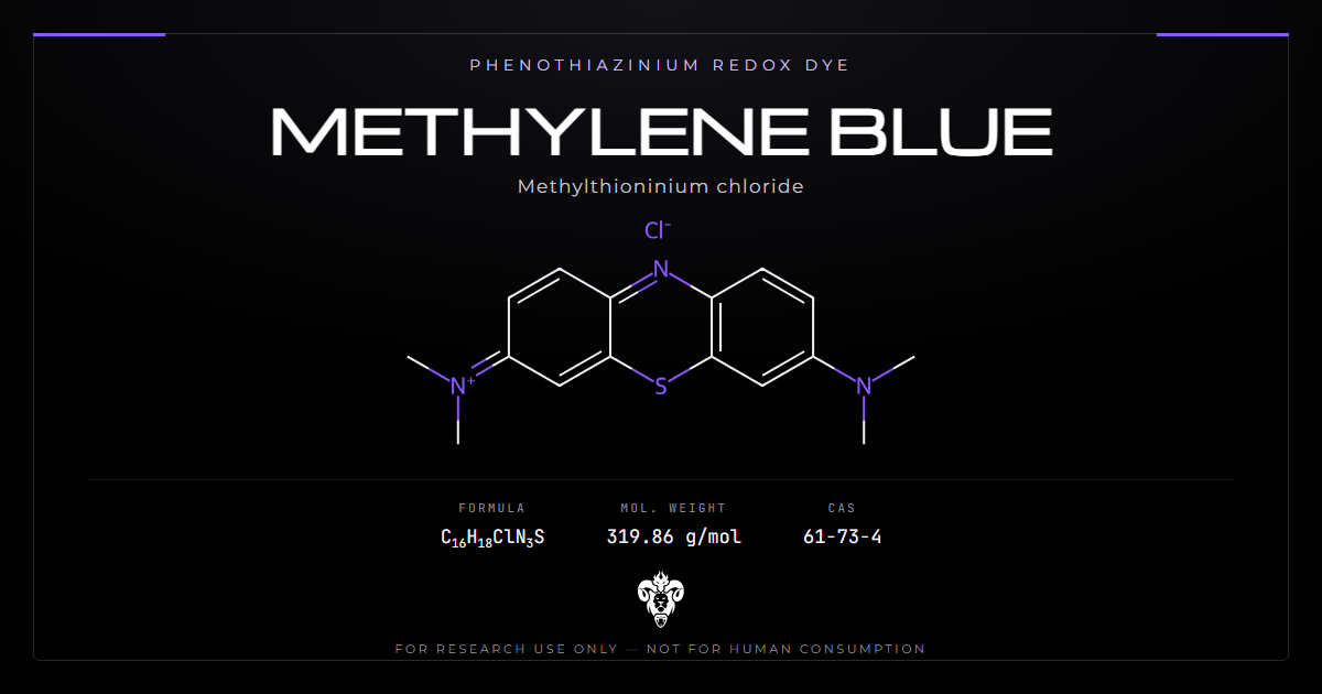
The compound is methylthioninium chloride. A tricyclic aromatic core carries a ring sulfur and a ring nitrogen. Dimethylamino groups sit at positions 3 and 7. A delocalised positive charge balances against chloride.
| Property | Value |
|---|---|
| Systematic name | [7-(dimethylamino)phenothiazin-3-ylidene]-dimethylazanium chloride |
| Common names | Methylene blue, methylthioninium chloride, MB |
| Colour Index name | C.I. Basic Blue 9 |
| Molecular formula | C16H18ClN3S |
| Molecular weight | 319.85 g/mol |
| Monoisotopic mass | 319.0910 Da |
| CAS number | 61-73-4 |
| PubChem CID | 6099 |
| InChIKey | CXKWCBBOMKCUKX-UHFFFAOYSA-M |
| Chemical class | Phenothiazinium redox dye |
| Reduced form | Leucomethylthioninium (colourless) |
| Common hydrate | Trihydrate |
The catalogue carries it as methylene blue.
The name covers two oxidation states
Methylene blue is one half of a redox couple. The oxidised form is the blue cation everyone recognises. Accept two electrons and a proton and it becomes leucomethylthioninium. That leuco form is colourless.
The colour change is the assay. A solution that has lost its blue has been reduced. The two forms differ in pharmacology, not merely in appearance. The tau programme discussed below develops the reduced form as a separate chemical entity [12][13].
Redox potential sits near 11 mV. That value lands inside the respiratory chain. NADH can reduce the dye. Cytochrome c can oxidise the leuco form. Those two facts are the electron-shuttle claim.
Dye grade and chemical grade are not the same specification
Basic Blue 9 has served as an industrial textile and biological stain for over a century. Most of the world’s production is made to that standard. Dye-grade material carries heavy metals. Arsenic, aluminium, cadmium, chromium, lead and zinc appear at levels acceptable for staining fabric.
Those metals are identity and purity questions. A certificate that reports only an HPLC area percent has not answered them. Elemental analysis belongs on the sheet next to the organic purity. A lot that matches C16H18ClN3S and fails metals is still a failed lot.
Azure A, azure B and azure C are demethylated relatives. They arise as manufacturing impurities and as metabolites. Azure B also inhibits MAO [9]. Its presence changes the pharmacology rather than merely diluting it.
Where the compound came from
Heinrich Caro synthesised methylene blue at BASF in 1876 as a textile dye. Its move into biology came through staining. The molecule binds some tissues and organisms and not others. That selectivity suggested selective chemistry.
Paul Ehrlich pursued that inference in the 1890s. The structural legacy outlived the early biological uses. Methylene blue is the first lead structure of the phenothiazines [2]. The ring system later produced a large set of related dyes and drugs.
That history explains an awkward feature of the present literature. The compound predates modern target-driven design. Mechanistic work arrived roughly a century after the first biological uses. Wen and Poteet are late papers on an old reagent (PMID 21454572, PMID 23118969).
The redox couple is the whole mechanism
Most of what methylene blue does follows from one property. It cycles between two oxidation states at a potential that sits inside the respiratory chain.
Alternative electron transfer
Wen and colleagues demonstrated the mechanism directly (PMID 21454572). Methylene blue accepts electrons from NADH and transfers them to cytochrome c. The route bypasses complexes I and III [1].
The control experiment is the convincing part. They synthesised a derivative with the redox centre disabled by N-acetylation. That analogue had no effect on mitochondrial complex activities. The redox cycling is the mechanism, not a correlate of it.
Poteet and colleagues extended this across seven structurally related compounds [2]. Compounds without side chains at positions 3 and 7 carry different redox potentials and cannot enhance electron transfer. They do retain direct antioxidant activity. Substituting a side chain at the ring nitrogen cut protective potency by three orders of magnitude.
What that does to oxygen consumption
Rerouting electrons past two complexes has measurable consequences. Cellular oxygen consumption rises. Anaerobic glycolysis falls [1]. Complex IV expression and activity increase [2].
The selectivity is informative. Methylene blue protected cultured neurons against glutamate, iodoacetic acid and rotenone. Those three insults involve mitochondrial impairment. Against direct oxidative stress from glucose oxidase it gave no protection at all [2]. It is not a general antioxidant. It is specifically a bypass for a blocked chain.
Kondak later found hydromethylthionine inside isolated brain mitochondria from tau-transgenic mice [13]. Respiration stayed intact in wild-type animals and rose in one transgenic line. That paper is about the reduced entity, not the blue chloride salt. Shared redox chemistry is not shared lot identity.
Biphasic redox in cells and animals
This is the part that gets lost when the compound is discussed casually.
Nanomolar in cells
The protective concentrations in the cell work are low nanomolar (PMID 21454572) [1]. That sits well below the concentrations producing visible blue in tissue.
Above that window the molecule stops shuttling electrons. It becomes a redox cycler generating the species it otherwise mitigates. The same chemistry that makes it useful at low concentration makes it harmful at high concentration.
Write the concentration next to every claim. A result at 10 nM and a result at 10 uM are different experiments. They are not two points on one straight line.
Why animal groups converge on the low end
Lu and Li both used 0.5 mg/kg/day in rats, in unrelated models, arriving at it independently [6][8]. Park used a four-day pretreatment in mice [4]. Those are small animal exposures.
Groups working on the electron-carrier mechanism converge on the low end. Groups that exploit direct chemical reduction of haemoglobin sit at a different concentration band. Those are two pharmacologies reached with one molecule. Reading across between them is the most common error in discussions of this compound.
Indexed exposure-response papers in people exist [11][12]. Human dose ladders sit outside this profile. The cell and animal inversion is the fact that organises the bench work.
Preclinical record
The animal and cell work is consistent in mechanism and consistent in dose. Almost all of it uses sub-milligram-per-kilogram exposures or nanomolar baths.
| Study | Model | Exposure | Principal finding |
|---|---|---|---|
| Wen 2011 [1] | Rotenone, rat | Low dose | Striatal dopamine depletion largely rescued |
| Wen 2011 [1] | Transient focal ischaemia | Low dose | Reperfusion damage reduced |
| Poteet 2012 [2] | HT-22 cells | Nanomolar | Protection via electron bypass; none against glucose oxidase |
| Park 2014 [4] | Noise, BALB/c mice | 4 days pretreatment | Threshold shift and outer hair cell death reduced |
| Burelle 2015 [5] | Leigh syndrome fibroblasts | In vitro | Protective against palmitate-plus-lactate death |
| Lu 2015 [6] | Global cerebral ischaemia, rat | 0.5 mg/kg/day, 7 days | CA1 survival improved; CCO and ATP restored |
| Li 2016 [8] | Streptozotocin ICV, rat | 0.5 mg/kg/day, 7 days | Memory deficit reduced; CCO and ATP increased |
| Kondak 2023 [13] | Tau-transgenic mice | Hydromethylthionine | Respiration intact in wild type; rose in one line |
The Leigh syndrome result deserves attention
Burelle and colleagues tested ten interventions in fibroblasts from patients with the French-Canadian variant of Leigh syndrome (PMID 25835550) [5]. That variant is a cytochrome c oxidase deficiency.
Compounds promoting flux through the electron transport chain, methylene blue and dinitrophenol among them, were protective. Three antioxidants, idebenone, N-acetylcysteine and resveratrol, made cell death worse.
That result separates two things routinely conflated. Methylene blue is not protective because it mops up radicals. In this model the antioxidants failed and the electron carriers worked.
Convergence on complex IV
Four independent groups working in different tissues report one downstream marker. Cytochrome c oxidase activity is preserved or restored, with ATP following [4][6] and [8]. Cochlea, hippocampus after ischaemia, and hippocampus after streptozotocin are unrelated preparations converging on one measurement.
Park showed that complex IV activity stayed up in the protected cochlea [4]. Lu restored cytochrome c oxidase and ATP in CA1 after global ischaemia [6]. Li restored the same pair after intracerebroventricular streptozotocin [8]. A laboratory that wants a positive control can pick that enzyme assay.
MAO-A inhibition chemistry
Methylene blue is a potent monoamine oxidase A inhibitor. That is a binding fact, not a slogan.
Numbers on the enzyme
Delport and colleagues measured an IC50 of 0.07 uM against human MAO-A, specific for that isoform over MAO-B [9]. Several structural analogues were more potent still. Cresyl violet sat at 0.0037 uM. Nile blue sat at 0.0077 uM.
Azure B, the demethylated impurity, also inhibits MAO [9]. A lot that fails the azure check is a mixed MAO reagent. HPLC with photodiode array detection separates the family. The visible spectrum differs enough to be diagnostic.
Existing cited plasma arithmetic
Gillman reviewed the interaction literature (PMID 20142303) [3]. An intravenous dose of 0.75 mg/kg gave a peak plasma concentration of 500 ng/ml, roughly 1.6 uM. That published peak sits more than twenty times the MAO-A IC50.
Indexed case series exist in that paper. Human toxicity scores sit outside this profile. The enzyme number and the published plasma figure are the parts that travel to a bench notebook.
Laboratory work that combines this compound with serotonergic agents faces a characterised, potent enzyme interaction. State the MAO-A IC50 next to the bath concentration. A nanomolar electron-shuttle experiment and a micromolar MAO experiment are different designs.
Haemoglobin redox and NADPH
NADPH reduces methylene blue to the leuco form. The leuco form can donate electrons to ferric haemoglobin. At higher concentration the oxidised dye can oxidise haemoglobin directly. Direction depends on concentration and on the reducing system.
The concentration paradox is chemistry
That pair of reactions is the clearest illustration of the inverted redox. It is also why the compound cannot be discussed sensibly without a stated concentration. The same vial can reduce or oxidise a haem protein. The difference is how much is in the cuvette and how much NADPH is present.
Sherwood and colleagues discuss this chemistry in an indexed case report on rasburicase-associated methaemoglobin [7]. Human case management sits outside this profile. The NADPH dependence does not.
G6PD is an NADPH supply question
Reduction to the leuco form depends on NADPH. Red cells generate it through the pentose phosphate pathway, using glucose-6-phosphate dehydrogenase.
A cell that lacks G6PD cannot supply enough NADPH. The dye stays oxidised. It then acts as an oxidant stress on a system already short of reducing power. That is a genotype-dependent reversal of the redox direction. It is a cell-biochemistry fact. It is not a dosing instruction.
Fibroblast and erythrocyte assays can test the point. A G6PD-null preparation is a different reagent condition from a wild-type one. Write the genotype next to the oxygen-consumption trace.
Hydromethylthionine is a different lot
Methylene blue inhibits pathological aggregation of tau in published models. That observation led to a development programme that is worth separating from the parent salt.
A reduced, stabilised entity
Hydromethylthionine is the reduced, stabilised form, developed as a distinct entity. Indexed phase 3 papers exist in Alzheimer’s disease and in frontotemporal dementia [11][12]. Those human endpoints sit outside this profile.
Wischik and colleagues set out the rationale for a separate oral programme (PMID 36281683) [12]. Shiells and colleagues published an exposure-response analysis on that entity [11]. Do not transfer those trial numbers onto a vial labelled methylene blue.
The compound sold as methylene blue is not hydromethylthionine. They share a redox couple and a tau-aggregation literature. They are handled, stabilised and named differently. Intact mass and oxidation state decide which one is in the vial.
Mouse mitochondria on the reduced form
Kondak and colleagues examined mitochondrial consequences in tau-transgenic mice (PMID 37445987) [13]. The assay found hydromethylthionine inside isolated brain mitochondria. Rivastigmine and memantine both lowered mitochondrial respiration in that comparison. The reduced dye did not.
Read that as a hypothesis about the leuco form in mouse brain mitochondria. It does not identify an unknown blue salt. It does not license a claim about people.
Limits of the published record
Four gaps sit between the published record and the claims that circulate around it.
Population and certainty
Indexed surgical and dementia papers exist [10][11] and [12]. Human pain scores, human atrophy scores and human dose ladders are out of scope here. A 2022 design paper and a 2020 exposure-response paper stay in the list so a reader can find them.
Which oxidation state was measured?
The blue cation and the leuco form are different species. A methods section that names methylene blue and then quotes a hydromethylthionine mass has switched lots. Absorbance near 665 nm reports the oxidised form. A colourless solution has been reduced, or degraded, or both.
Which grade was in the vial?
No published study has compared dye-grade against chemical-grade material on a biological endpoint. The metals distinction is well established chemically and untested pharmacologically. A certificate without elemental analysis has not closed the question.
What this profile will not do
It will not quote human milligram-per-day ladders or human optimum windows. Those sentences turn a research article into a use document. The papers remain cited [10][11] and [12].
Four questions to ask of any methylene blue result
Most disagreement about this compound traces to comparing figures that were never comparable.
Which structure was measured?
Formula C16H18ClN3S, mass 319.85 Da, CAS 61-73-4, CID 6099, InChIKey CXKWCBBOMKCUKX-UHFFFAOYSA-M. A lot that fails those checks is not methylene blue.
Which oxidation state?
Blue cation or leuco form. Absorbance at 665 nm with a shoulder near 610 nm is the oxidised dye. A clear solution is not.
Which concentration band?
Nanomolar electron shuttle, micromolar MAO block, and high-concentration oxidant chemistry are different experiments [1][3] and [9]. State the bath or the mg/kg.
Is the comparator the same backbone?
Hydromethylthionine, azure B and cresyl violet share a phenothiazine story and do not share a certificate. A result that names “a redox dye” without naming the lot is not usable.
Verifying research material
Purity for this compound means more than a chromatographic percentage.
Identity
Intact mass 319.85 Da average, 319.09 monoisotopic. The chloride counterion is part of the formula. A free-base mass without chloride is a different line on the sheet.
Visible spectrum is the cheapest identity check available. The oxidised form absorbs strongly near 665 nm with a shoulder around 610 nm. A reduced leuco form does not absorb there at all. A sample whose absorbance ratio is off, or whose solution is not the expected blue, is either reduced, degraded or not what the label says.
Water content matters for weighing. The trihydrate is the common form. Treating it as anhydrous introduces a systematic error of roughly 14 percent in molar terms.
Heavy metals and related dyes
Heavy metals are the specific purity concern, since dye-grade material is the default global production. A certificate should report elemental analysis, not just organic purity. Arsenic, lead, cadmium, chromium, aluminium and zinc are the names to look for.
Related dyes are the second concern. Azure A, azure B and azure C demethylate off the parent. HPLC with photodiode array detection separates them. Azure B also inhibits MAO [9], so the impurity is a second active species.
Handling
Store the solid cold, dry and dark. Reconstitute close to the point of use.
Storage guidance is a house recommendation. Analytical documentation is per-lot release testing.
Light and reductants bleach the blue. That bleach can be reversible reduction or irreversible degradation. A UV-visible scan before use is cheaper than a failed assay. Plastic binds the cation. Dilute solutions lose material to tube walls. Aliquot on reconstitution rather than sampling one vial repeatedly.
Kimera publishes third-party certificates of analysis for every lot in its COA database. Laboratories source methylene blue as a redox-active reference material. It often sits alongside BAM15 and DADA, which reach mitochondrial bioenergetics by unrelated routes, or emoxypine succinate where a direct antioxidant is the comparator. Related chemistry appears in the nootropics category.
Common questions about methylene blue
Why would a lower concentration outperform a higher one? The redox cycling that donates electrons at low concentration generates reactive species at high concentration. Cell work measured this in the nanomolar window [1].
Does the blue colour indicate activity? It indicates oxidation state, which is not the same thing. The blue cation is the oxidised form. The reduced leuco form is colourless. A solution that has gone clear has been reduced rather than spoiled, though it may also have degraded.
Is it an antioxidant? Not primarily. It gave no protection against direct oxidative stress from glucose oxidase, while protecting against insults that impair mitochondrial complexes [2]. In Leigh syndrome fibroblasts, three genuine antioxidants worsened cell death where methylene blue helped [5].
What is the MAO-A number? IC50 0.07 uM, selective over MAO-B [9]. Gillman published a 500 ng/ml peak after 0.75 mg/kg intravenous, about 1.6 uM [3].
Why does G6PD status reverse the redox? Reduction to the leuco form needs NADPH from the pentose phosphate pathway. Without G6PD that supply fails. The compound then stays oxidised and adds to the oxidant burden instead of relieving it [7].
Is dye-grade material the same compound? The same molecule, a different specification. Dye-grade production meets a standard that permits heavy metal content irrelevant to staining fabric.
Do the hydromethylthionine papers apply to it? No. Those papers used the stabilised reduced form, developed as a separate entity [12][13]. Indexed human trials on that entity exist [11][12]. This profile does not quote them as methylene blue results.
Summary of the evidence
Write the name, the mass and the redox couple on the first line of a notebook page. Everything else in this profile is a check on those facts.
Identity: methylthioninium chloride, C16H18ClN3S, 319.85 g/mol, CAS 61-73-4. A phenothiazinium redox dye, also catalogued as C.I. Basic Blue 9. PubChem CID 6099 and InChIKey CXKWCBBOMKCUKX-UHFFFAOYSA-M close the record.
Mechanism: an alternative electron carrier. It accepts electrons from NADH and donates them to cytochrome c, bypassing complexes I and III [1]. Disabling the redox centre abolishes the effect [1].
Selectivity: protective against mitochondrial insults, not against direct oxidative stress [2]. Electron carriers helped where antioxidants harmed in a cytochrome c oxidase deficiency model [5].
Convergent marker: cytochrome c oxidase activity and ATP preserved across cochlear, ischaemic and streptozotocin models [4][6] and [8].
Concentration-response: biphasic, nanomolar in cells [1]. Animal groups working the electron-carrier mechanism converge near 0.5 mg/kg/day [6][8].
Enzyme: MAO-A inhibition, IC50 0.07 uM [9]. Published peak plasma after 0.75 mg/kg intravenous is 500 ng/ml [3].
Related entity: hydromethylthionine, the reduced form. Mouse mitochondrial data exist [13]. Indexed human papers exist [11][12]. Not the same material.
Limits: human efficacy, human dose and human adverse-event figures are out of scope here. Those papers remain in the reference list.
Status: supplied for laboratory research use only.
References
- Wen Y, Li W, Poteet EC, et al. Alternative mitochondrial electron transfer as a novel strategy for neuroprotection. J Biol Chem. 2011;286(18):16504-16515. PMID 21454572. DOI
- Poteet E, Winters A, Yan LJ, et al. Neuroprotective actions of methylene blue and its derivatives. PLoS One. 2012;7(10):e48279. PMID 23118969. DOI
- Gillman PK. CNS toxicity involving methylene blue: the exemplar for understanding and predicting drug interactions that precipitate serotonin toxicity. J Psychopharmacol. 2011;25(3):429-436. PMID 20142303. DOI
- Park JS, Jou I, Park SM. Attenuation of noise-induced hearing loss using methylene blue. Cell Death Dis. 2014;5(4):e1200. PMID 24763057. DOI
- Burelle Y, Bemeur C, Rivard ME, et al. Mitochondrial vulnerability and increased susceptibility to nutrient-induced cytotoxicity in fibroblasts from Leigh syndrome French Canadian patients. PLoS One. 2015;10(3):e0120767. PMID 25835550. DOI
- Lu Q, Tucker D, Dong Y, Zhao N, Zhang Q. Neuroprotective and functional improvement effects of methylene blue in global cerebral ischemia. Mol Neurobiol. 2016;53(8):5344-5355. PMID 26433378. DOI
- Sherwood GB, Paschal RD, Adamski J. Rasburicase-induced methemoglobinemia: case report, literature review, and proposed treatment algorithm. Clin Case Rep. 2016;4(4):315-319. PMID 27099716. DOI
- Li L, Qin L, Lu HL, Li PJ, Song YJ, Yang RL. Methylene blue improves streptozotocin-induced memory deficit by restoring mitochondrial function in rats. Brain Res. 2017;1657:208-214. PMID 28034723. DOI
- Delport A, Harvey BH, Petzer A, Petzer JP. The monoamine oxidase inhibition properties of selected structural analogues of methylene blue. Toxicol Appl Pharmacol. 2017;325:1-8. PMID 28377303. DOI
- Farrokhi MR, Lotfi M, Masoudi MS, Gholami M. Effects of methylene blue on postoperative low-back pain and functional outcomes after lumbar open discectomy: a triple-blind, randomized placebo-controlled trial. J Neurosurg Spine. 2016;24(1):7-15. PMID 26360148. DOI
- Shiells H, Schelter BO, Bentham P, et al. Concentration-dependent activity of hydromethylthionine on clinical decline and brain atrophy in a randomized controlled trial in behavioral variant frontotemporal dementia. J Alzheimers Dis. 2020;75(2):501-519. PMID 32280089. DOI
- Wischik CM, Bentham P, Gauthier S, Miller S, Kook K, Schelter BO. Oral tau aggregation inhibitor for Alzheimer’s disease: design, progress and basis for selection of the 16 mg/day dose in a phase 3, randomized, placebo-controlled trial of hydromethylthionine mesylate. J Prev Alzheimers Dis. 2022;9(4):780-790. PMID 36281683. DOI
- Kondak C, Leith M, Baddeley TC, et al. Mitochondrial effects of hydromethylthionine, rivastigmine and memantine in tau-transgenic mice. Int J Mol Sci. 2023;24(13):10810. PMID 37445987. DOI
Methylene blue is sold for laboratory research use only. Not for human consumption, nor medical, veterinary, or household uses.
Literature retrieved from PubMed.