
Everything below reports findings from cell cultures, isolated tissue and animals. This material is for research use only, not for human or veterinary use.
Give MOTS-c to a mouse on a high-fat diet and it prevents the insulin resistance and the obesity that diet would otherwise produce [1]. That result launched the field in 2015 and it has held up.
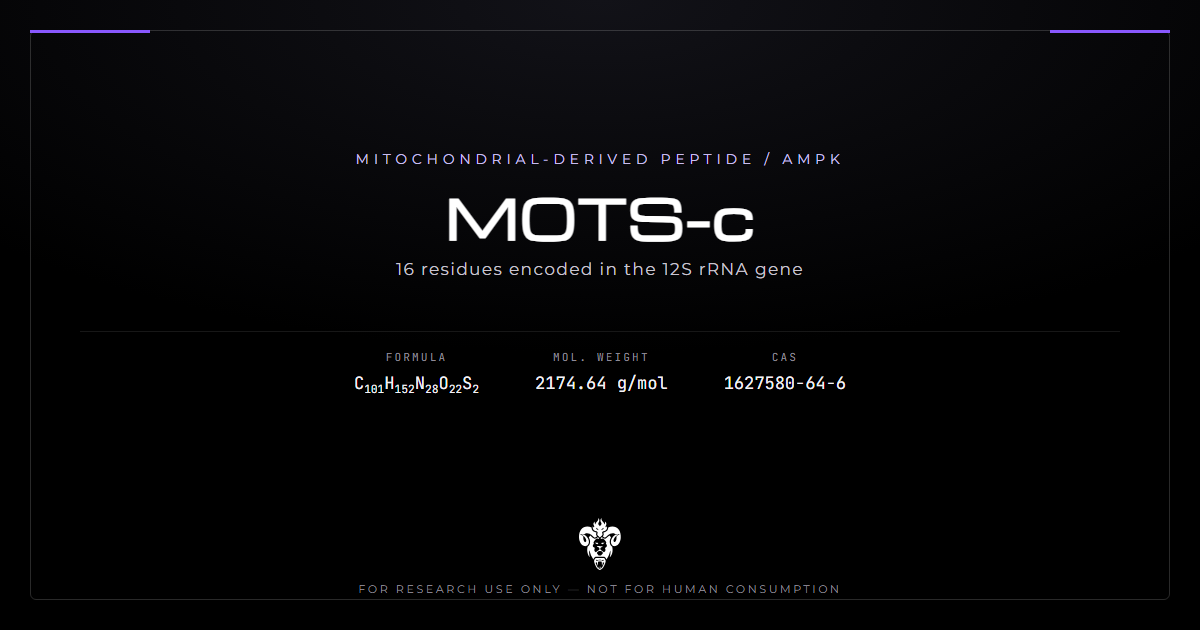
The peptide is sixteen residues long. The instructions live in mitochondrial DNA, not in the nucleus. That location is the identity fact that matters most.
Indexed measurement papers in people exist. Those circulating-level and disease endpoints sit outside this profile. The useful laboratory questions are sequence, the folate-cycle route, and what mouse and cell systems measured.
Chemical identity
Sixteen residues, and the striking thing is where the instructions for them live.
| Property | Value |
|---|---|
| Sequence | Met-Arg-Trp-Gln-Glu-Met-Gly-Tyr-Ile-Phe-Tyr-Pro-Arg-Lys-Leu-Arg |
| Common names | MOTS-c, mitochondrial ORF of the 12S rRNA type-c |
| Molecular formula | C101H152N28O22S2 |
| Molecular weight | 2174.6 |
| CAS | 1627580-64-6 |
| PubChem CID | 146675088 |
| InChIKey | WYTHCOXVWRKRAH-LOKRTKBUSA-N |
| Methionines | Two, positions 1 and 6 |
| Basic residues | Arg, Arg, Arg, Lys |
Kimera supplies the material as MOTS-c.
Encoded in mitochondrial DNA, not nuclear
The nuclear genome encodes nearly every peptide in this catalogue. MOTS-c is not. It comes from a short open reading frame inside the mitochondrial 12S ribosomal RNA gene [1].
That is a strange place to keep a gene. The mitochondrial genome is tiny, roughly 16.5 kilobases, and it was long assumed to encode only the respiratory chain subunits, tRNAs and rRNAs it obviously needs.
Reading the sequence
Two methionines sit at positions 1 and 6. Both are oxidation liabilities. Three arginines and one lysine make the chain strongly basic. Tryptophan at position 3 gives a real 280 nm chromophore.
Proline at position 12 breaks helix. The C-terminal Arg-Lys-Leu-Arg stretch is the charge cluster. A deletion in that cluster changes both mass and ion-exchange behaviour.
It was found by looking for a second one
Humanin came first, a signalling peptide encoded in a short ORF in mitochondrial DNA. Its discovery raised an obvious question: if one exists, are there others?
Humanin is a different sequence, a different mass, and a different experimental literature. Shared organelle is not shared pharmacology. Keep the two vials on separate labels.
MOTS-c is the answer to that question [1]. The search was deliberate, which is worth knowing because it means the finding was not a fluke of annotation.
The mechanism is unusually specific
Most peptides in this catalogue act on a receptor. MOTS-c acts on a metabolic pathway.
The folate cycle route
Its cellular action inhibits the folate cycle and the de novo purine biosynthesis tethered to it, and that inhibition leads to AMPK activation [1]. Skeletal muscle appears to be the primary target organ.
This is a metabolic-intermediate mechanism rather than a receptor-binding one. That distinction matters for experimental design: there is no obvious antagonist to use as a control, and no binding assay to run.
AMPK is the convergence point
AMPK activation connects the peptide to a large literature on metabolic regulation. Reviewers therefore group the compound with agents like AICAR.
Sharing an endpoint is not sharing a mechanism. Those compounds reach AMPK by different routes, and read-across between them is unreliable.
A laboratory that wants an AMPK control should still name the route. AICAR is a nucleotide analogue. MOTS-c is a 16-residue peptide that starves the folate cycle first [1].
Folate-cycle inhibition also starves one-carbon units used in methylation. That is a second reason to read more than AMPK. A methyl-donor panel and a purine panel answer different questions about the same peptide.
What to measure in a cell experiment
Folate-cycle intermediates and purine nucleotides are the proximal readouts. AMPK phosphorylation is downstream. A study that reports only AMPK has skipped the claimed mechanism.
Skeletal muscle cells are the obvious first system [1]. A line that does not run a folate cycle will not reproduce the founding result.
What the mouse data showed
The founding study is clean, and it bounds its own claims carefully.
MOTS-c treatment in mice prevented age-dependent insulin resistance, prevented high-fat-diet-induced insulin resistance, and prevented diet-induced obesity [1].
Note the verb. Prevented, in animals treated alongside the insult. That is a different claim from reversing established disease, and the distinction survives into every later discussion of the compound.
The age-dependent result deserves its own line. Insulin resistance that develops with age in an untreated mouse is a slower, less artificial model than diet-induced disease, and preventing it is the more interesting half of that paper.
None of it involved a deficient animal. The mice made their own peptide throughout, and the treatment added to a normal baseline rather than correcting a shortfall.
Pairing MOTS-c with humanin later attenuated atrial fibrillation readouts in a fibrosis and mitochondrial-dysfunction model [9]. That design cannot assign the result to either peptide alone.
The founding paper also mapped the open reading frame and the protein product in the same study [1]. Sequence, mechanism and mouse phenotype arriving together is unusual. It is also why later papers keep citing that one source for three different claims. Treat each claim separately when you design a follow-up.
Indexed measurement papers
Several indexed papers measure circulating or tissue MOTS-c in people rather than administer the synthetic peptide. Those human endpoints sit outside this profile.
| Study | What was measured | Why it sits outside |
|---|---|---|
| Obesity cohort [2] | Circulating peptide | Human association, not administration |
| Lipid-infusion cohort [3] | Plasma peptide after a metabolic insult | Human association |
| Stromal cells [4] | Expression and exogenous peptide in culture | Cell work, kept below |
| Metformin biomarker [5] | Circulating peptide during another agent | Association |
| Alzheimer pairing [6] | Expression with telomere length | Association |
| Diabetes review [7] | Circulating peptide in disease | Association |
| Polymorphism [8] | m.1382A>C against a clinical label | Association |
The useful laboratory lesson from that table is simple. Most of the later literature measures the endogenous peptide. Measurement is not administration.
Tissue expression can disagree with plasma
The stromal-cell paper found basal MOTS-c expression lower in mesenchymal stromal cells from obese donors than lean ones [4]. Circulating papers point the other way [2]. Those two need not contradict each other if tissue releases the peptide under stress. Nobody has demonstrated that release step in a closed system.
Why compartment matters in the laboratory
A compound whose endogenous level moves with metabolic state is not obviously a simple deficiency reagent. Two readings stay open in animals. Either added peptide still does work on top of a stress response, which is what the founding mouse paper tested [1]. Or the rise is reporting on a problem elsewhere. Mouse prevention data cannot close that argument by themselves.
The stromal cell result is the sharpest caution
That 2026 study did more than measure. It gave MOTS-c to cells and looked at what happened [4].
Exogenous MOTS-c restored intracellular levels in obese-donor cells and activated metabolic signalling, which is the expected result. It also blunted reparative function, which is not.
The design extended to stenotic mouse kidneys, testing whether pre-treated obese stromal cells repaired tissue better. The headline says they did not.
One study, and it deserves replication rather than deference. It is also the only published work that gave the compound to primary human cells and measured something other than a metabolic marker.
Where else it appears
The literature has expanded quickly, and most of it measures MOTS-c rather than administering it.
As a biomarker
Investigators have examined circulating levels in several disease cohorts [5][6] and [7]. Another group tested a polymorphism, m.1382A>C [8].
Biomarker studies are cheaper than intervention studies, which is why they dominate. They also cannot establish that changing the level changes anything. Their human endpoints stay outside this profile.
The atrial fibrillation work is the exception
That study administered the peptide alongside humanin rather than measuring it, and reported attenuated fibrosis and mitochondrial dysfunction [9]. It belongs in the intervention column.
Pairing the two mitochondrial-derived peptides is a sensible design, given they were discovered the same way and share an organelle. It also means the study cannot attribute its result to either one alone.
Reviews
Reviews cover its relationship to stress, metabolism and ageing [10], to exercise-induced muscle-fat crosstalk [11], and to mitochondrial microproteins more broadly [12]. Reviews are maps. They are not mouse data.
A review that cites the founding paper for mechanism, mouse phenotype and mitochondrial origin is citing one study three times [1][10]. That is fine if the reader sees it. It is not three independent confirmations.
What being mitochondrially encoded actually implies
The location of the gene is not trivia. It carries three consequences that a nuclear-encoded peptide would not have.
Maternal inheritance
Mitochondrial DNA passes down the maternal line only. So any sequence variant in this gene, and any functional consequence of one, follows a different inheritance pattern from every nuclear gene.
That is why a polymorphism such as m.1382A>C attracts attention [8]. Variants in mitochondrial DNA cluster into haplogroups that track ancient population history, which makes them unusually tractable for association studies and unusually easy to confound with ancestry.
Heteroplasmy
A cell carries hundreds to thousands of mitochondrial genome copies, and they need not be identical. The proportion carrying a variant can differ between tissues in the same animal and can shift over a lifetime.
Nuclear genes do not behave like that. For this peptide it means “genotype” is a fuzzier concept than usual, and a blood sample may not report what muscle carries.
Copy number is a variable
Mitochondrial DNA copy number varies with tissue, age, exercise and disease. If expression scales with copy number even loosely, then the amount of peptide a tissue makes depends on something that moves for reasons unrelated to the peptide itself.
That is one plausible route to a circulating-versus-tissue discrepancy, and nobody has ruled it in or out.
The exercise thread
Reviews repeatedly connect this peptide to exercise, and the connection is worth stating precisely because it gets overstated.
What the reviews say
Work on exercise-induced muscle-fat crosstalk treats the peptide as one mediator among several [11]. The broader reviews position it within stress, metabolism and ageing [10].
Skeletal muscle is the primary target organ identified in the founding work [1], so a muscle-exercise link is mechanistically reasonable rather than invented.
What it does not establish
Exercise raising a molecule does not make that molecule the reason exercise works. Dozens of things rise with exercise, and most of them are downstream consequences rather than causes.
A level that moves with metabolic state may be reporting on that state rather than driving it. This peptide gives a clear example of that problem, which is part of why it is worth reading carefully.
How to read a MOTS-c study
Four questions, and the second is the one most reviews skip.
Measured or administered?
Most later MOTS-c papers measure endogenous levels [2][3] and [5][6]. Those describe an association. Only the administration studies say anything about what the compound does [1][4] and [9].
Which direction, and in what compartment?
Published work can put circulating and tissue levels in opposite directions [2][4]. A paper calling the peptide reduced or elevated without naming the compartment has left out half the claim.
Prevention or reversal?
The founding mouse work prevented metabolic disease in animals treated alongside the insult [1]. Extending that to reversal is an assumption, not a finding.
Was reparative function measured?
The one study that looked found metabolic signalling and repair moving in opposite directions [4]. Metabolic markers alone may miss that.
Verifying research material
MOTS-c is a defined chemical entity with published identifiers, so verification is arithmetic rather than judgement. Batch documentation sits on the certificates of analysis page.
Identity
Formula C101H152N28O22S2, molecular weight 2174.6, InChIKey WYTHCOXVWRKRAH-LOKRTKBUSA-N. The PubChem name lookup resolves to a stereo-defined record here.
Note the two sulfurs. Both methionines, at positions 1 and 6, and they are the compound’s main analytical liability.
Methionine oxidation is the check that matters
Methionine oxidises readily to the sulfoxide, adding 16 daltons per residue. With two methionines a preparation can sit at 2174.6, 2190.6 or 2206.6, and the oxidised forms are not the compound every published study used.
This is not a hypothetical. An N-terminal methionine oxidises especially readily, and position 1 here is exactly that. Ask whether the certificate reports oxidised species separately or only a single purity figure.
Tryptophan can oxidise too. Watch plus-16 and plus-32 in the mass channel, and do not assume a clean parent ion.
Handling follows from that
Oxygen and light both drive the oxidation. Store the lyophilised solid at minus 20 degrees Celsius, dark, with minimal headspace in the vial. Reconstituted solutions degrade faster than the solid, so treat them as short-lived.
Storage guidance is a house recommendation. Analytical documentation is per-lot release testing.
There is no cysteine, so disulfide scrambling is not a concern, which distinguishes it from many peptides of similar size. Compare with Epithalon, a much shorter peptide with a very different stability profile.
Aliquot on reconstitution. Repeated freeze-thaw of a methionine-rich peptide is how a 2174.6 lot becomes a 2190.6 lot without anyone changing the label.
Peptide content is not chromatographic purity. Lyophilised material carries counterions and water. A vial labelled by weight holds less peptide than that mass, and acetate or trifluoroacetate belongs on the certificate next to the free-base mass.
The three arginines make trifluoroacetate a likely leftover from purification. Residual TFA carries its own activity in some cell assays. Ask for it, or exchange it, before a low-concentration experiment.
Common questions about MOTS-c
Identity and origin
What does the name mean? Mitochondrial open reading frame of the 12S rRNA type-c. It describes exactly where the gene sits.
Is it encoded in mitochondrial DNA? Yes, in a short open reading frame within the 12S rRNA gene [1]. That is unusual and it is the compound’s defining feature.
How was it found? By deliberate search. Humanin had already shown that mitochondrial DNA encodes signalling peptides, and MOTS-c came from looking for another [1].
How does it work? It inhibits the folate cycle and the de novo purine synthesis attached to it, which activates AMPK. Skeletal muscle appears to be the main target [1].
Evidence
What did the mouse work show? Prevention of age-dependent and diet-induced insulin resistance, and of diet-induced obesity [1].
Do circulating papers exist? Yes. Indexed measurement papers exist [2][3]. Their human endpoints sit outside this profile.
Is there a negative finding in cells? One. In human stromal cells, exogenous MOTS-c activated metabolic signalling but blunted reparative function [4].
What is the main handling risk? Methionine oxidation. Two methionines, 16 daltons each, one of them N-terminal.
Why does mitochondrial encoding matter? Three reasons: maternal inheritance, heteroplasmy, and copy number that varies with tissue and age. None of those apply to a nuclear-encoded peptide.
Is it exercise-related? Reviews connect it to exercise-induced muscle-fat crosstalk [11]. That a molecule rises with exercise does not make it the reason exercise works.
Has anyone published administration studies in people? Indexed measurement papers exist. Administration endpoints in people sit outside this profile.
Handling and verification
What mass should a batch return? 2174.6. Readings at 2190.6 or 2206.6 indicate one or both methionines oxidised.
Does it contain cysteine? No, so disulfide scrambling is not a concern.
How should a certificate report purity? With oxidised species separated out, not folded into a single number. Ask specifically, because a single figure hides the failure mode this sequence is most prone to.
Does PubChem resolve it correctly by name? Yes, to a stereo-defined record. That is not true of every peptide in this catalogue.
Summary of the evidence
Strongest evidence: the founding characterisation [1], which identified the open reading frame, the sequence, the folate-cycle mechanism, the AMPK endpoint and the mouse metabolic phenotype in one paper. That is a lot of independent claims in a single study, and the mechanism has held.
Also solid: the cell study that saw metabolic signalling and reparative function move in opposite directions [4]. Pairing with humanin in an atrial-fibrillation model is intervention work that cannot split the two peptides [9].
Indexed measurement papers in people exist. Those endpoints sit outside this profile.
Read plainly, MOTS-c is a well-characterised discovery with a specified mechanism and an unresolved interpretation. The mouse data says giving it helps prevent a diet or age insult. Endogenous levels move with metabolic state in other papers. Nobody has closed that gap in a single design.
For laboratory purposes that ambiguity is workable. The identity is settled, the mechanism is specified, and the folate-cycle route gives a concrete thing to measure.
The rest of the peptide literature sits in the peptides category.
Status: supplied for laboratory research use only.
References
- Lee C, Zeng J, Drew BG, Sallam T, Martin-Montalvo A, Wan J, Kim SJ, Mehta H, et al. The mitochondrial-derived peptide MOTS-c promotes metabolic homeostasis and reduces obesity and insulin resistance. Cell Metab. 2015;21(3):443-54. PMID 25738459. DOI
- Yoon SH, Yuan F, Zhu X, Tang H, Abdurakhimova D, Krier J, Eirin A, Lerman A, et al. Systemic MOTS-c levels are increased in adults with obesity in association with metabolic dysregulation and remain unchanged after weight loss. J Clin Transl Endocrinol. 2026;43:100429. PMID 41551324. DOI
- Ramanjaneya M, Jerobin J, Bettahi I, Bensila M, Aye M, Siveen KS, Sathyapalan T, Skarulis M, et al. Lipids and insulin regulate mitochondrial-derived peptide (MOTS-c) in PCOS and healthy subjects. Clin Endocrinol (Oxf). 2019;91(2):278-287. PMID 31066084. DOI
- Xing L, Lu B, Zhu X, Al Saeedi M, Lerman A, Eirin A, Cohen P, Lerman LO. Mitochondrial-derived peptide MOTS-c activates metabolic signaling but blunts reparative function in human mesenchymal stromal cells. Inflamm Regen. 2026;. PMID 42324588. DOI
- Cuyàs E, Verdura S, Martin-Castillo B, Menendez JA. Circulating levels of MOTS-c in patients with breast cancer treated with metformin. Aging (Albany NY). 2022;15(4):892-897. PMID 36490309. DOI
- Rodríguez-Esparragón F, Cazorla-Rivero SE, Torrealba E, Cánovas-Molina Á, González-Hernández AN, Martín-Alfaro R, Afonso-Medina MP, Martínez de Saavedra-Álvarez MT, et al. Insights into the Biomarker Potential of Humanin and Mots-c Expression and Telomere Length in Alzheimer’s Disease. Int J Mol Sci. 2025;26(22). PMID 41303353. DOI
- Fang T, Han JC, Taberner A, Pham T. MOTS-c in type 2 diabetes mellitus: From risk factors to cardiac complications and potential treatment. Life Sci. 2025;382:124009. PMID 41083123. DOI
- Filibeli BE, Dedemoglu F, Garipçin P, Bulut S, Başok Bİ, Kizildağ S, Dündar B, Çatli G. Are serum MOTS-c levels and MOTS-c m.1382A>C polymorphism related to polycystic ovary syndrome?. Arch Endocrinol Metab. 2026;70(3):e260031. PMID 41945630. DOI
- Liao Y, Xu J, Jiao Y, Sun X, Gao M, Ding Y, Cai D, Shen Y, et al. Humanin and MOTS-c Attenuate Atrial Fibrillation by Suppressing Fibrosis and Mitochondrial Dysfunction. Biomedicines. 2026;14(5). PMID 42193373. DOI
- Wan W, Zhang L, Lin Y, Rao X, Wang X, Hua F, Ying J. Mitochondria-derived peptide MOTS-c: effects and mechanisms related to stress, metabolism and aging. J Transl Med. 2023;21(1):36. PMID 36670507. DOI
- Tero-Vescan A, Degens H, Matsakas A, Ștefănescu R, Ősz BE, Slevin M. Exercise-Induced Muscle-Fat Crosstalk: Molecular Mediators and Their Pharmacological Modulation for the Maintenance of Metabolic Flexibility in Aging. Pharmaceuticals (Basel). 2025;18(8). PMID 40872612. DOI
- Patel K, Soni R, Shah J. The unexplored Nexus: Mitochondria derived microproteins and Parkinson’s disease. Pathol Res Pract. 2025;273:156136. PMID 40694987. DOI
MOTS-c is sold for laboratory research use only. Not for human consumption, nor medical, veterinary, or household uses.