
Everything below reports findings from cell cultures, isolated tissue and animals. This material is for research use only, not for human or veterinary use.
Roxadustat makes a cell behave as though oxygen were short. It blocks the enzymes that mark hypoxia-inducible factor for destruction. The factor survives, translocates, and switches on a coordinated gene set.
That chemistry is a 2-oxoglutarate competition, not a glycoprotein injection. The useful laboratory questions are identity, enzyme logic, and what animal systems actually measured.
A development programme and several indexed clinical papers exist. Those human endpoints sit outside the scope of this profile.
Chemical identity of Roxadustat
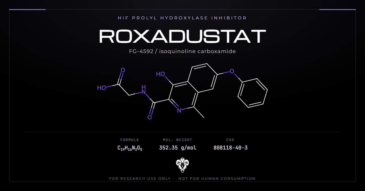
Roxadustat is an isoquinoline carboxamide. Development codes include FG-4592 and ASP-1517. PubChem lists CID 11256664.
| Property | Value |
|---|---|
| Common names | Roxadustat, FG-4592, ASP-1517 |
| Systematic | 2-[(4-hydroxy-1-methyl-7-phenoxyisoquinoline-3-carbonyl)amino]acetic acid |
| Molecular formula | C19H16N2O5 |
| Molecular weight | 352.34 g/mol |
| Monoisotopic mass | 352.1059 Da |
| CAS | 808118-40-3 |
| PubChem CID | 11256664 |
| InChIKey | YOZBGTLTNGAVFU-UHFFFAOYSA-N |
| Class | Isoquinoline carboxamide, HIF prolyl hydroxylase inhibitor |
| Stereocentres | None |
| Appearance | White to pale green solid |
The catalogue carries it as Roxadustat FG-4592.
Reading the structure
An isoquinoline core carries a phenoxy substituent, a methyl group and a phenol. A glycine amide hangs off the carboxamide. That glycinamide is the business end. It mimics 2-oxoglutarate, the co-substrate the prolyl hydroxylases need, and competes for the same site.
The compound is small and not a peptide. A 352 Da acid is a straightforward substrate for glucuronidation and for renal handling. It behaves nothing like a glycoprotein hormone in an assay. That is why the detection literature on Roxadustat reads like a small-molecule problem rather than a biologics problem, and why the numbers involved are picograms per millilitre [9].
The InChIKey carries the UHFFFAOYSA block. There are no defined stereocentres. Stereochemical verification is not the concern here that it is for peptides.
What the formula decides
C19H16N2O5 at 352.34 Da separates this lot from vadadustat, daprodustat and molidustat. Those neighbours hit the same enzyme class on different scaffolds. Shared target language is not shared mass.
A certificate that quotes a different monoisotopic mass is not Roxadustat, whatever the label says. Write 352.1059 Da next to the name.
HIF-PHD inhibition chemistry
Prolyl hydroxylase domain enzymes tag HIF-alpha for degradation when oxygen is plentiful. Inhibit them and the alpha subunit survives.
Competitive with 2-oxoglutarate
Del Balzo and colleagues measured recombinant full-length human HIF-PH enzymes with a 14C-labelled 2-oxoglutarate decarboxylation assay (PMID 32487538, DOI).
In the presence of 100 uM 2-oxoglutarate, 1 uM Fe2+ and 1 mM ascorbate, Roxadustat IC50 values were 1.04 uM at PHD1, 1.74 uM at PHD2 and 0.36 uM at PHD3. Dependence of PHD2 activity on 2-oxoglutarate at several inhibitor concentrations gave invariable Vmax and variable Km. That is competitive kinetics. The Ki at PHD2 was 20 nM.
Factor inhibiting HIF, another 2-oxoglutarate dioxygenase, gave an IC50 above 150 uM under the same conditions. The PHD panel is the target. FIH is not.
Treat those figures as assay numbers, not as a certificate. A laboratory that wants the trio for a given vial runs the enzymes.
How the hydroxylases mark HIF
PHD enzymes are iron-dependent dioxygenases. They use one oxygen atom to hydroxylate a proline on HIF-alpha. They use the other to decarboxylate 2-oxoglutarate to succinate. Oxygen, iron, ascorbate and the alpha-keto acid all have to be present. Remove any one and the mark fails.
Two prolines on HIF-1a carry the mark in the usual textbook map. Hydroxylated HIF-alpha then binds the von Hippel-Lindau ubiquitin ligase. The proteasome clears the subunit within minutes when oxygen is plentiful. Block the hydroxylase and that clock stops. The alpha subunit accumulates. It partners with HIF-beta. The heterodimer binds hypoxia-response elements.
Roxadustat does not copy a hypoxia gas mix. It copies the missing co-substrate. The glycinamide occupies the 2-oxoglutarate site. Oxygen can still be present. The enzyme still cannot complete the cycle. That is why the literature calls the state a transient pseudo-hypoxia [1]. The gas mix has not changed. The tag has.
Write the co-substrate concentration next to every IC50. Del Balzo used 100 uM 2-oxoglutarate. A screen that uses a different load will move the number. Competitive inhibitors look weaker when the co-substrate is high.
More than one gene
Locatelli and colleagues set out the pathway map (PMID 28118622) [1]. HIF drives erythropoietin. It also moves iron-handling genes through hepcidin and related nodes. A ligand that stabilises HIF therefore hits a programme, not a single hormone.
That coordination is the design argument against supplying a glycoprotein and leaving iron handling untouched. Whether a given lot engages both arms is an assay question. Hepcidin protein, ferroportin transcript, and erythropoietin protein are different measurements. A cell study that reports one of them has not reported the others.
Vadadustat and daprodustat occupy the same enzyme class [7]. Isoform rank order is not identical across the class. Do not transfer a PHD3-preferring profile onto a Roxadustat vial.
Animal and cell readouts
Nonclinical work is where the enzyme pair becomes a phenotype.
Cells, rats and monkeys in the nonclinical package
The same JPET file shows HIF-1a and HIF-2a protein rising in cells (PMID 32487538). Erythropoietin production rose even in the presence of EPO-suppressing inflammatory cytokines.
Intermittent oral administration in healthy rats and cynomolgus monkeys raised circulating erythropoietin, reticulocytes and haemoglobin in a dose-dependent manner. A five-sixths nephrectomy rat model and a peptidoglycan-polysaccharide inflammation model both moved those markers. The inflammation model also moved hepcidin.
Those are animal systems. They describe the mechanism. They do not set a human use.
Adenine-diet rats
Gao and colleagues gave adenine-diet CKD rats Roxadustat with and without a polysaccharide iron complex (PMID 40050784) [10].
The combination raised haemoglobin faster than either alone. The compound also lowered hepcidin, interleukin-6, tumour necrosis factor alpha, interleukin-1 beta and HMGB1. Masson staining and expression of alpha-smooth muscle actin and fibronectin both pointed to less renal fibrosis.
Those are rat data on an adenine model. Read them as mechanism, not as a transfer claim.
| Study | System | Exposure | Readout |
|---|---|---|---|
| Del Balzo 2020 | Recombinant PHD1/2/3 | Enzyme assay | IC50 1.04 / 1.74 / 0.36 uM; PHD2 Ki 20 nM |
| Del Balzo 2020 | Cells | Bath | HIF-1a and HIF-2a protein up |
| Del Balzo 2020 | Rat, monkey | Oral, intermittent | EPO, reticulocytes, haemoglobin up |
| Del Balzo 2020 | 5/6 nephrectomy rat | Oral | Anaemia markers moved |
| Del Balzo 2020 | PG-PS rat | Oral | Hepcidin down |
| Gao 2025 [10] | Adenine CKD rat | Oral, plus iron complex | Hb faster in combo; fibrosis markers down |
| Hansson 2017 [2] | Microsomes, fungus, hepatocytes | In vitro | Metabolite map depends on the model |
| Mathew 2021 [4] | Thoroughbred horse | Oral | 13 urinary metabolites |
Metabolite maps are a property of the model
Hansson and colleagues investigated the compound across five in vitro systems and one human doping-control sample (PMID 27918992) [2].
Twelve metabolites appeared in total. Human and equine liver microsomes each gave one monohydroxylated metabolite. The fungus Cunninghamella elegans gave eleven. Human hepatocytes and equine liver S9 fraction gave none at all. In the human urine sample, exactly one metabolite appeared, a direct glucuronide.
Mathew and colleagues then dosed thoroughbred horses orally and detected 13 urinary metabolites (PMID 33569900) [4]. Seven were phase I, one phase II and five conjugates of phase I products. Hydroxylation dominated, with a glucuronide as the major phase II product.
The lesson generalises. A metabolite map is a property of the model as much as of the molecule. Choosing hepatocytes over microsomes over a fungus changes the answer by an order of magnitude. A methods section that names “the Roxadustat metabolites” without naming the system has not named a result.
Those maps are also the impurity library for a chromatogram. Hydroxylated and glucuronidated species are the peaks a purity method should already know how to find [2][4].
A fungus that produces eleven products is a metabolite factory, not a proof that people make eleven. A hepatocyte dish that produces none is not a proof that the molecule is inert. It is a proof that that dish did not turn it over. Match the system to the question. Identity work wants the parent. Pathway work wants the enzyme. Excretion work wants the matrix that the method will actually see.
Detection windows and existing cited PK
Anti-doping laboratories, not nephrology clinics, produced the most unusual kinetic number in this file.
Months, not hours
Sobolevsky and colleagues report detection in urine for eight months after a single dose (PMID 39350653) [9]. Concentrations by then sit below 10 pg/mL. The authors describe the terminal excretion kinetics as unusually prolonged and tell testing authorities to account for it.
That profile mirrors what other small-molecule files in this catalogue have already shown at picogram sensitivity. Detection windows measured in months rather than days are a recurring feature of modern assays, not a unique magic of this scaffold.
Indexed clinical papers that discuss regular treatment schedules exist [5][6] and [9]. Human milligram ladders sit outside this profile. The eight-month single-dose window is the cited kinetic fact that belongs on an analytical method.
Cross-contamination at picogram sensitivity
Some adverse analytical findings were attributed to contaminated dietary supplements [9]. At picogram sensitivities, cross-contamination in a shared workspace becomes a real analytical risk rather than a theoretical one. Segregate weighing. Use dedicated glassware.
A 352 Da carboxylic acid sticks to surfaces. A later blank that prints a picogram peak has not proved a dosed animal. It has proved a dirty bench.
The rest of the class, as chemistry
Roxadustat is not alone. Three HIF prolyl hydroxylase inhibitors went through extensive published programmes: Roxadustat, vadadustat and daprodustat [7].
Shared target, different scaffolds
Every member competes with 2-oxoglutarate at PHD enzymes, raises HIF-alpha, and can be given orally in animal work. One further shared property appears in review: inflammation influences the class less than it influences injected erythropoiesis-stimulating agents, at least on the hepcidin argument [7].
That last point is a mechanism claim. Inflammation drives hepcidin. Hepcidin locks up iron. A mechanism that lowers hepcidin should be less vulnerable to that failure mode. The rat data support the hepcidin part of the chain directly [10]. Whether that theoretical advantage produces a better animal outcome in a new model is a new experiment.
Locatelli and Del Vecchio review the class (PMID 36041790) [7]. Indexed human comparisons sit in that paper. This profile does not quote them as a treatment contest.
Do not transfer isoform rank
If the compounds shared a clean class effect, one failing in one system would be surprising. If they do not, results from one agent do not transfer to another. Roxadustat’s file has to stand on its own enzyme and animal data. A paper that names “a HIF-PHI” without naming the lot is not usable.
ITPP works on the other side of the oxygen problem, shifting haemoglobin oxygen release rather than HIF stability. TND1128 and SLU-PP-332 sit in adjacent mitochondrial and metabolic work. Keep the names on separate labels.
Limits of the published record
Four gaps sit between the published record and the claims that circulate around it.
Population and certainty
Pooled phase 3 analyses, switching studies, left-ventricular papers and safety meta-analyses exist. Human haemoglobin scores, human adverse-event rates and approval labels sit outside this profile. A 2025 complementary-medicine review of renal anaemia sits in the list as a map [11]. Use it for bibliography. Do not use it as a use document.
Which arm is being credited
Erythropoietin protein, hepcidin protein and fibrosis histology are different readouts [1][10]. A paper that reports haemoglobin in a rat and then claims iron mobilisation has skipped a measurement.
Which molecule was in the vial
A 352 Da isoquinoline acid is easy to confuse with another HIF-PHI. Intact mass is the first check. A name on a label is not.
What this profile will not do
It will not quote human dose ladders, human adverse-event rates, or approval labels. Those sentences turn a research article into a use document. The papers remain cited so a reader can find them [12].
Four questions to ask of any Roxadustat result
Most disagreement about this compound traces to comparing figures that were never comparable.
Which structure was measured?
Formula C19H16N2O5, mass 352.34 Da, CAS 808118-40-3, CID 11256664, InChIKey YOZBGTLTNGAVFU-UHFFFAOYSA-N. A lot that fails those checks is not Roxadustat.
Which enzyme assay?
State the PHD isoform, the 2-oxoglutarate concentration, and whether FIH was read. Dual or triple inhibition is a measured set, not a class nickname.
Which animal model?
Five-sixths nephrectomy, PG-PS inflammation, and adenine-diet CKD are different systems [10]. Read-across between them is a claim that needs its own experiment.
Is the comparator the same scaffold?
Vadadustat, daprodustat and injectable erythropoietin proteins share a conversation and do not share a certificate [7]. A result that names “a HIF-PHI” without naming the lot is not usable.
Verifying research material
The compound is a defined small molecule with published identifiers, so verification is measurement. Batch documentation sits on the certificates of analysis page.
Identity
Formula C19H16N2O5, molecular weight 352.34, CAS 808118-40-3, InChIKey YOZBGTLTNGAVFU-UHFFFAOYSA-N. Electrospray in negative mode should return [M-H]- near 351.10. Positive mode should return [M+H]+ near 353.11.
Both the isoquinoline core and the phenoxy ether absorb strongly in the ultraviolet. That absorbance is the cheapest identity check after mass. A pale green tint on the solid is common and is not, by itself, a fail. A brown oil is.
The 4-hydroxyisoquinoline can tautomerise on paper. The InChI still resolves to one connectivity. A vendor drawing that moves the phenol to another ring carbon is a different compound. Compare the InChIKey, not the cartoon.
Purity and related substances
Chromatography with diode-array detection separates the compound and its related substances readily. The published metabolite maps give a reference for what hydroxylated and glucuronidated species look like on a chromatogram [2][4].
Phenol regioisomers and a missing glycine are the synthetic impurities to hunt first. Each has a defined mass offset. Drop the glycine and the parent falls by 75 Da. Move the phenoxy and the UV spectrum shifts while the mass stays put. A single area-percent on a poor column will hide both.
Counterion and residual solvent belong on the sheet. A carboxylic acid isolated from an amide coupling often carries acetate, trifluoroacetate or sodium. Peptide-content logic does not apply here, but salt form still changes the mass of a weighed vial. Do not infer the salt from the free-acid formula.
What a doping method already solved
Hansson and Mathew ran high-resolution mass methods built to find picogram residues [2][4]. Those traces are a gift to a release lab. They already list the hydroxylated masses and the glucuronide. A purity method that cannot see those peaks is not finished.
Sobolevsky then showed how long a urine method can still print a peak [9]. That paper is a warning about persistence and about dirty supplements. It is also a reminder that this acid survives in matrices that chew up peptides. Plan the blank series accordingly.
Handling
Store the solid cold, dry and dark. Reconstitute close to the point of use.
Storage guidance is a house recommendation. Analytical documentation is per-lot release testing.
The carboxylic acid makes the solid hygroscopic relative to a neutral analogue. Weigh quickly. Cap the vial. Picogram-sensitive work needs dedicated glassware [9]. A shared spatula is a contamination route, not a convenience.
Laboratories source Roxadustat as a HIF prolyl hydroxylase inhibitor reference. Related work appears in the metabolic category.
Common questions about Roxadustat
What is Roxadustat? An isoquinoline glycinamide, C19H16N2O5, 352.34 Da, CAS 808118-40-3. It is a HIF prolyl hydroxylase inhibitor.
How does the enzyme block work? The glycinamide mimics 2-oxoglutarate. Binding is competitive at PHD2 with a Ki of 20 nM (PMID 32487538).
What are the PHD IC50 values? About 1.04 uM at PHD1, 1.74 uM at PHD2 and 0.36 uM at PHD3 in the decarboxylation assay. FIH sat above 150 uM.
Does this page report human outcomes? No. Indexed clinical papers are listed so they can be found. This profile stops at chemistry, enzyme logic and animal systems.
What did the adenine-diet rats show? Faster haemoglobin rise when an iron complex sat alongside, plus lower hepcidin and lower fibrosis markers [10].
How long can a urine method still see it? Eight months after a single dose, at concentrations below 10 pg/mL [9].
Why do metabolite counts disagree? Hansson found one product in human microsomes and eleven in a fungus [2]. Mathew found thirteen in horse urine [4]. The model writes the map.
Summary of the evidence
Write the name, the mass and the three PHD isoforms on the first line of a notebook page. Everything else in this profile is a check on those facts.
Identity: C19H16N2O5, 352.34 Da, CAS 808118-40-3, CID 11256664, InChIKey YOZBGTLTNGAVFU-UHFFFAOYSA-N. A certificate that omits intact mass is not finished.
Roxadustat is the long name for that lot. FG-4592 and ASP-1517 are the same chain under development codes. Do not treat a code as a second compound.
Design: a 2-oxoglutarate-competitive isoquinoline that stabilises HIF-alpha [1]. Del Balzo supplies the enzyme numbers and the rat and monkey readouts.
Animal evidence: nephrectomy and inflammation models in the nonclinical package, plus adenine-diet rats with an iron-complex arm [10]. Hepcidin and fibrosis markers moved in that last system.
Analytical evidence: model-dependent metabolite maps [2][4] and an eight-month urinary detection window after a single dose [9].
Limits: human efficacy, human dose and human adverse-event figures are out of scope here. Those papers remain in the reference list [3][8].
Status: supplied for laboratory research use only.
References
- Locatelli F, Fishbane S, Block GA, Macdougall IC. Targeting hypoxia-inducible factors for the treatment of anemia in chronic kidney disease patients. Am J Nephrol. 2017;45(3):187-199. PMID 28118622. DOI
- Hansson A, Thevis M, Cox H, Miller G, Eichner D, Bondesson U, Hedeland M. Investigation of the metabolites of the HIF stabilizer FG-4592 (roxadustat) in five different in vitro models and in a human doping control sample using high resolution mass spectrometry. J Pharm Biomed Anal. 2017;134:228-236. PMID 27918992. DOI
- Ogawa C, Tsuchiya K, Tomosugi N, Maeda K. Hypoxia-inducible factor prolyl hydroxylase domain inhibitor may maintain hemoglobin synthesis at lower serum ferritin and transferrin saturation levels than darbepoetin alfa. PLoS One. 2021;16(6):e0252439. PMID 34143801. DOI
- Mathew B, Philip M, Perwad Z, Karatt TK, Caveney MR, Subhahar MB, Karakka Kal AK. Identification of hypoxia-inducible factor (HIF) stabilizer roxadustat and its possible metabolites in thoroughbred horses for doping control. Drug Test Anal. 2021;13(6):1203-1215. PMID 33569900. DOI
- Barratt J, Sulowicz W, Schomig M, Esposito C, Reusch M, Young J, Csiky B. Efficacy and cardiovascular safety of roxadustat in dialysis-dependent chronic kidney disease: pooled analysis of four phase 3 studies. Adv Ther. 2021;38(10):5345-5360. PMID 34523074. DOI
- Zhu XW, Zhang CX, Xu TH, Jiang GN, Yao L. Efficacy of roxadustat in treatment of peritoneal dialysis patients with renal anaemia. World J Clin Cases. 2021;9(26):7682-7692. PMID 34621819. DOI
- Locatelli F, Del Vecchio L. Hypoxia-inducible factor-prolyl hydroxyl domain inhibitors: from theoretical superiority to clinical noninferiority compared with current ESAs? J Am Soc Nephrol. 2022;33(11):1966-1979. PMID 36041790. DOI
- Tan W, Wang X, Sun Y, Wang X, He J, Zhong L, Jiang X, Sun Y, Tian E, Li Z, He L, Hao Y, Tang B, Hua W, Ma X, Yang J. Roxadustat reduces left ventricular mass index compared to rHuEPO in haemodialysis patients in a randomized controlled trial. J Intern Med. 2024;295(5):620-633. PMID 38343089. DOI
- Sobolevsky T, Fedoruk M, Dellanna F, Geyer H, Ahrens B, Thevis M. Long-term excretion of roxadustat in urine. Drug Test Anal. 2025;17(7):1088-1092. PMID 39350653. DOI
- Gao Z, Gao Y, Wang Q, Wang Q, Lu P, Lv H, Xue H, Ma X, Li S, Hu Z. Study on HIF-PHI combined with iron supplement in treatment of renal anemia in rats. BMC Nephrol. 2025;26(1):125. PMID 40050784. DOI
- Lu CM, Hsu YH, Lin IH, Kuo KL, Liao JF, Huang HF, Lu PH. Conventional and complementary alternative medicine therapies for renal anemia: a literature review. Front Endocrinol (Lausanne). 2025;15:1342873. PMID 39911241. DOI
- Martinez-Miguel P, Fernandez-Anton E, Rodriguez-Puyol D, de Abajo FJ, Lopez Ongil S. Safety of roxadustat in chronic kidney disease patients: an updated systematic review and meta-analysis. Pharmaceuticals (Basel). 2025;18(10):1566. PMID 41155680. DOI
Roxadustat is sold for laboratory research use only. Not for human consumption, nor medical, veterinary, or household uses.
Literature retrieved from PubMed.